

In situ Hybridization (ISH) and Quantum Dots (QD) of miRNAs
miRNA的原位杂交(ISH)和量子点(QD)


发布: 2017年02月20日第7卷第4期 DOI: 10.21769/BioProtoc.2138 浏览次数: 11636
评审: HongLok LungChiara AmbrogioAnonymous reviewer(s)
参见作者原研究论文
The authors used this protocol in:

May 2015
Advertisement


Abstract
miRNA are short non-coding RNA which inhibit translation of mRNA. miRNA regulate several cellular processes. Certain miRNA are known to induce oncogenesis. miRNA can be measured by real-time PCR and be imaged using a combination of in situ hybridization (ISH) and quantum dots (QD). The advantage of using quantum dots is that several miRNA can be simultaneously measured using multiplexed QD. Additionally, miRNA can be visualized in different regions of the tissue. Since miRNA are biomarkers of various disease states, miRNA can be visualized and quantitated in tissue sections for diagnostic and prognostic purposes. Here we describe ISH-QD analysis of tissue sections. Tissue sections from xenografts or clinical specimens are used. These are deparaffinized, treated with Proteinase K and hybridized with a biotin-probe to specific to the miRNA. The in situ hybridization is performed by labeling the biotin-probes and followed by labeling with streptavidin tagged quantum dots. Image acquisition of the quantum dots is performed and analyzed for the miRNA expression levels. Combining ISH and QD gives a powerful tool to detect miRNA in different cells of the tissue.
Keywords: miRNA (miRNA)Background
miRNAs can be easily detected by quantitative real-time PCR or Northern blotting. However, imaging miRNAs has been challenging. Recent advances in quantum dots imaging have made it possible to determine expression of miRNAs in tissues. Using this process, miRNAs can be visualized in different compartments of a tissue, such as tumor, stroma, immune cells, etc. Additionally, miRNAs can be multiplexed to determine co-localization of miRNAs which mediate specific processes, in different cellular regions. Different tissues can be used for ISH-QD such as tissues from xenografts or human clinical samples. Tissues from animal studies are formalin fixed and paraffin embedded. These tissues were used for ISH-QD analysis (Figure 1). 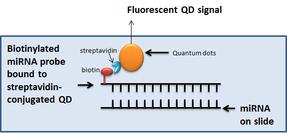
Figure 1. In situ hybridization coupled to Quantum dots labelling for visualization of miRNA. In the ISH-QD protocol, miRNA are detected on formalin fixed paraffin embedded tissue sections using biotinylated miRNA probes binding to streptavidin-conjugated QDs. The specific QD gives a specific fluorescent signal which is quantified using the Inform v1.3 software.
Materials and Reagents
- Gloves
- Tissue slides
- Superfrost plus slides
- Coverslips
- LNATM Scramble-miR probe (Exiqon, catalog number: 699004-370 )
- LNATM miR-409-3p 5’ biotin labeled (Exiqon, catalog number: 610701-370 )
- LNATM miR-409-5p 5’ biotin labeled (Exiqon, catalog number: 615615-370 )
- RNaseZap (Thermo Fisher Scientific, AmbionTM)
- Xylene
- Ethanol
- Sterile PBS (pH 7)
- Nail polish
- Horse serum (Vector Laboratories, catalog number: S-2000 )
- Streptavidin block reagent (Thermo Fisher Scientific, InvitrogenTM)
- Streptavidin conjugated QDs 625 nm (Thermo Fisher Scientific, Molecular ProbesTM, catalog number: A10196 ) (1 µm stock from Invitrogen)
- Streptavidin conjugated QDs 565 nm (Thermo Fisher Scientific, Molecular ProbesTM, catalog number: Q10131MP ) (1 µm stock from Invitrogen)
- 6% IgG-free, protease free BSA (Jackson ImmunoResearch, catalog number: 001-000-162 )
- BSA
- 0.4% Triton X-100
- 0.1% Tween-20
- Mounting media
- 4’6-diamidino-2-phenylindole (DAPI) (Vector Laboratories)
- 1 M Tris-HCl (pH 7.4)
- 0.5 M EDTA
- NaCl
- RNase free Milli-Q water, autoclaved (all solutions prepared in this water)
- Exiqon microRNA ISH buffer set and Proteinase K (Exiqon, catalog number: 90000 )
- 20x SSC, pH 7.0
- Proteinase K stock (see Recipes)
- Proteinase K buffer (see Recipes)
- Proteinase K solution (15 µg/ml) (see Recipes)
- SSC solutions (see Recipes)
- Hybridization mix (see Recipes)
- Streptavidin blocking solution (see Recipes)
- QD solutions (see Recipes)
- PBS-T (see Recipes)
Equipment
- Autoclave
- Hybridizer (DAKO Statspin Hybridizer)
- Glass cutter
- Slide rack and glass jars
- PAP pen or ImmEdge pen (Vector Laboratories, catalog number: H-4000 )
- Water bath
- CRi multi-spectral camera with built-in Nuance software and inForm software (PerkinElmer, Waltham, MA)
Software
- Nuance v3.1 software
- Inform v1.3 software
- Graphpad Prism software
Procedure
文章信息
版权信息
© 2017 The Authors; exclusive licensee Bio-protocol LLC.
如何引用
Josson, S., Gururajan, M. and Chung, L. W. (2017). In situ Hybridization (ISH) and Quantum Dots (QD) of miRNAs. Bio-protocol 7(4): e2138. DOI: 10.21769/BioProtoc.2138.
分类
癌症生物学 > 通用技术 > 生物化学试验 > RNA
生物化学 > RNA > miRNA标记
您对这篇实验方法有问题吗?
在此处发布您的问题,我们将邀请本文作者来回答。同时,我们会将您的问题发布到Bio-protocol Exchange,以便寻求社区成员的帮助。