

Protocol for Murine/Mouse Platelets Isolation and Their Reintroduction in vivo
鼠/小鼠血小板分离及其在体内再导入实验方案


发布: 2017年02月20日第7卷第4期 DOI: 10.21769/BioProtoc.2132 浏览次数: 20475
评审: Lee-Hwa TaiShravani MukherjeeXiaoyi ZhengKate Hannan
参见作者原研究论文
The authors used this protocol in:
Mar 2012
Advertisement


Abstract
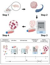
Platelets and coagulation have long been known to be essential for metastasis in experimental models. In order to study the interactions between tumor cells, platelets and endothelium, we have adapted methods used in coagulation research for the isolation of platelets and their reintroduction into mice. Anti-coagulated murine blood served as the source for platelets. Platelets were separated from other elements of the whole blood by centrifugation. Here the critical elements are first inhibition of coagulation and second isolation and maintenance of the platelets in the presence of inhibitors of platelet activation. We then used the vital dye PKH26 to fluorescently label the platelets. Infusion of these labelled platelets allows microscopic observation of the introduced platelets. After reintroduction, these platelets appear to function normally and comprise approximately 50% of the total platelets. Because they are fluorescently labelled, they can easily be identified. Finally it would be possible to use these methods for the determination of specific effects of altered gene expression in platelets by using platelets from genetically engineered mice. These methods have facilitated study of the interactions between platelets and tumor cells in tissue culture and in murine models. They would also be applicable to video microscopy. Here we provide details of the methods we have used for platelet isolation from mice and their staining for further microscopy and re-introduction into mice.
Keywords: Platelets (血小板)Background
Platelets are known to be essential for metastasis, but also to play roles during tumor growth not to mention clot formation. In order to readily identify and track platelets we developed the means for fluorescently labeling and reinfusing platelets. This allows them to be readily identified in tissues without immunostaining. Using these methods, we showed that interactions between tumor cells and platelets play key roles in survival of the tumour cells early during metastasis (Im et al., 2004; Gil-Bernabe et al., 2012). Platelets formed clots with tumor cells in the blood stream and this coagulation promoted spreading and subsequent retention of the tumor cells during lung metastasis (Im et al., 2004). Tissue factor expressed by tumor cells is capable of mediating clot formation with platelets and recruitment of macrophages (Gil-Bernabe et al., 2012). The interaction of platelets, thrombin and fibrin also have been reported to promote metastasis by generating epithelial-mesenchymal transition of the cancer cells and evasion from the immune system by protection from NK cells as well as secretion of pro-metastatic chemokines and cytokines (Labelle et al., 2011; Nieswandt et al., 1999; Palumbo et al., 2005 and 2007). Precise tracking of platelets will provide opportunities to uncover how tumor cells utilize the host for their survival.
Materials and Reagents
- Scalpel (Swann Morton, catalog number: 0208 )
- Syringe, 1 ml (BD, catalog number: 300013 )
- 15 ml conical bottom polypropylene tube (SARSTEDT, catalog number: 62.554.502 )
- 5 ml pipette
- Needle (27 G)
- Mice (4-6 weeks old, weight over 20 g, Charles Liver, UK)
- Isoflurane (Abbott, catalog number: 0044-5260-05 )
- EGTA (Sigma-Aldrich, catalog number: E3889 )
- PKH26 Kit (Sigma-Aldrich, catalog number: PKH26GL-1KT )
- Sodium chloride, NaCl (Thermo Fisher Scientific, Fisher Scientific, catalog number: 10378573 )
- Potassium chloride, KCl (Thermo Fisher Scientific, Fisher Scientific, catalog number: 10427460 )
- Sodium phosphate monobasic monohydrate, NaH2PO4·H2O (Sigma-Aldrich, catalog number: S9638 )
- HEPES (Sigma-Aldrich, catalog number: H3375 )
- Glucose (Thermo Fisher Scientific, GibcoTM, catalog number: 15023021 )
- Magnesium chloride, MgCl2 (Sigma-Aldrich, catalog number: M8266 )
- Trisodium citrate (Thermo Fisher Scientific, Fisher Scientific, catalog number: 10362234 )
- Citric acid (Sigma-Aldrich, catalog number: 251275 )
- Dextrose (Thermo Fisher Scientific, Fisher Scientific, catalog number: D16-1 )
- Prostaglandin E1 (Sigma-Aldrich, catalog number: P5515 )
- 100% ethanol
- Sodium bicarbonate, NaHCO3 (Thermo Fisher Scientific, Fisher Scientific, catalog number: S637-212 )
- Distilled water
- Bovine serum albumin, BSA (Sigma-Aldrich, catalog number: A2058 )
- Sodium bicarbonate, Na2HPO4 (Thermo Fisher Scientific, Fisher Scientific, catalog number: S374 )
- Sodium citrate (Sigma-Aldrich, catalog number: 71498 )
- Modified Tyrode’s calcium-free buffer (see Recipes)
- ACD buffer (see Recipes)
- Undiluted prostaglandin E1 solution
- Resuspension buffer (see Recipes)
- Washing buffer (see Recipes)
- Citrate-albumin buffer (see Recipes)
Equipment
- Coulter counter (CDC Technology, model: HEMAVET® 1500 )
- Centrifuge (Thermo Fisher Scientific, model: Jouan CR4i )
Procedure
文章信息
版权信息
© 2017 The Authors; exclusive licensee Bio-protocol LLC.
如何引用
Im, J. H. and Muschel, R. J. (2017). Protocol for Murine/Mouse Platelets Isolation and Their Reintroduction in vivo. Bio-protocol 7(4): e2132. DOI: 10.21769/BioProtoc.2132.
分类
癌症生物学 > 侵袭和转移 > 动物模型 > 细胞分离和培养
免疫学 > 动物模型 > 小鼠
细胞生物学 > 细胞分离和培养 > 细胞分离
您对这篇实验方法有问题吗?
在此处发布您的问题,我们将邀请本文作者来回答。同时,我们会将您的问题发布到Bio-protocol Exchange,以便寻求社区成员的帮助。