

Isolation of PBMCs Using Vacutainer® Cellular Preparation Tubes (CPTTM)
使用Vacutainer®细胞制备管(CPTTM)分离外周血单核细胞

发布: 2017年01月20日第7卷第2期 DOI: 10.21769/BioProtoc.2103 浏览次数: 38120
评审: Emmanuel ZavalzaMartin V KolevAnonymous reviewer(s)
参见作者原研究论文
The authors used this protocol in:
0 2014
Advertisement


Abstract
Peripheral blood mononuclear cell (PBMC) isolation is commonly done via density gradient centrifugation over Ficoll-Hypaque, a labor-intensive procedure that requires skilled technicians and can contribute to sample variability. Cellular Preparation Tubes (CPTs) are Vacutainer blood draw tubes that contain Ficoll-Hypaque and a gel plug that separates the Ficoll solution from the blood to be drawn. Once blood is drawn into CPTs, they can be centrifuged to separate the PBMC, then shipped (if desired) to a processing lab. The processing lab removes the PBMC from the upper compartment of the tube (above the gel plug), washes the PBMC, and can cryopreserve them using DMSO-containing media, as detailed in this protocol.
Keywords: PBMC (PBMC)Background
Isolation and cryopreservation of peripheral blood mononuclear cells (PBMC) is common practice in clinical studies that employ cellular immune assays. Cryopreservation allows for batching of samples, which is convenient and improves the comparability of data. Cryopreservation also allows cells to be stored for unknown future purposes. Because erythrocytes and granulocytes are much more fragile to freezing and thawing, PBMC isolation is a common prerequisite to cryopreservation of blood cells; and the most common method for PBMC isolation is density gradient centrifugation using Ficoll-Hypaque, a high molecular weight carbohydrate solution.
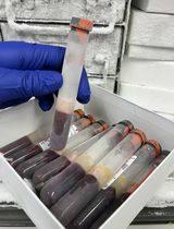
Cellular Preparation Tubes (CPTs), which contain Ficoll-Hypaque, simplify the standard procedure for density gradient centrifugation in two ways: (1) blood is collected into the same tube that is then used to isolate the PBMC; and (2) the tube is pre-loaded with Ficoll-Hypaque, which is separated by a gel plug so that it is not disturbed by the entry of blood into the tube. After blood draw, the tubes are centrifuged, and the PBMCs and plasma become separated from the erythrocytes and granulocytes by the gel plug (see Figure 1). This allows the spun tubes to be shipped, maintaining the PBMC in an isolated environment from the erythrocytes and granulocytes, which may improve their viability and function.
CPTs are ideal for use in studies which collect whole blood across multiple sites and ship to a central processing laboratory (Ruitenberg et al., 2006); this reduces variability in PBMC isolation between technicians. Studies have found no significant difference in PBMCs isolated using the CPT system or by traditional layover methods via density gradient separation (Corkum et al., 2015; Ruitenberg et al., 2006). While material cost can be high, CPTs reduce the length of processing time in addition to decreasing inconsistency between operators; both of these are key to reducing cost and increasing sample quality and consistency. Furthermore, when used with studies or sites which ship blood samples overnight, PBMCs from CPTs have a higher purity and less infiltrate from other (contaminating) cell types, such as red blood cells, in contrast to samples shipped in standard blood collection tubes over a 24-48 h period (Schlenke et al., 1998).
Figure 1. Empty CPT (left), after blood draw (middle), and after centrifugation (right). Location of gel plug and sample layers after centrifugation are shown.
Materials and Reagents
- 1.8 ml Nunc (or similar) cryovials
- BD Vactuainer® Mononuclear Cell Preparation Tubes (CPTTM), 8 ml, with sodium heparin (BD, catalog number: 362753 )
- P10 and P200 pipette tips (any vendor)
- P10 and P200 mechanical pipettors (any vendor)
- 50 ml Falcon (or similar) conical polypropylene tubes
- Serological pipettes (assorted volumes)
- BioCision CoolCell® (BioCision, catalog number: BCS172 ) – alcohol free controlled-rate freezing container
- Foam packing material and shipping containers
- Phosphate buffer saline (PBS),Ca2+ and Mg2+ free (e.g., Thermo Fisher Scientific, GibcoTM, catalog number: 10010-023 )
- Trypan blue (e.g., GE Healthcare, HycloneTM, catalog number: SV30084.01 or Sigma-Aldrich, catalog number: 93595 )
- Human AB Serum (e.g., Valley Biomedical, catalog number: HP1022 )
- Dimethyl sulfoxide (e.g., Sigma-Aldrich, catalog number: D8418 )
Equipment
- A centrifuge capable of reaching speeds of 1,800 x g (e.g., Beckman Coulter, model: Allegra X-14 Series )
- 50 ml conical adapters and buckets for centrifuge
- Pipette gun (any vendor)
- Automated cell counter or microscope and hemacytometer (any vendor)
Note: Cell counting methods vary in their throughput, cost, and flexibility for different cell types. For consistency, the same counting method should be used throughout a study. - Biosafety cabinet level A2 (BSC) (any vendor)
- -80 °C freezer (for initial cryopreservation) (any vendor)
- Liquid nitrogen (LN2) freezer (for long-term cryopreservation) (any vendor)
Note: LN2 systems range from small and simple to large and highly automated. Auto-filling and self-monitoring systems are recommended, especially for larger studies.
Procedure
文章信息
版权信息
© 2017 The Authors; exclusive licensee Bio-protocol LLC.
如何引用
Puleo, A., Carroll, C., Maecker, H. T. and Gupta, R. (2017). Isolation of PBMCs Using Vacutainer® Cellular Preparation Tubes (CPTTM). Bio-protocol 7(2): e2103. DOI: 10.21769/BioProtoc.2103.
分类
免疫学 > 免疫细胞分离 > 白细胞
细胞生物学 > 细胞分离和培养 > 细胞分离
您对这篇实验方法有问题吗?
在此处发布您的问题,我们将邀请本文作者来回答。同时,我们会将您的问题发布到Bio-protocol Exchange,以便寻求社区成员的帮助。