

Plant Tissue Trypan Blue Staining During Phytopathogen Infection
植物病原体感染期间的植物组织台盼蓝染色

发布: 2016年12月20日第6卷第24期 DOI: 10.21769/BioProtoc.2078 浏览次数: 34926
评审: Arsalan DaudiRupesh PaudyalRenate Weizbauer
参见作者原研究论文
The authors used this protocol in:
Jan 2002


Abstract
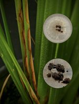
In this protocol plant tissue is stained with trypan blue dye allowing the researcher to visualize cell death. Specifically this method avoids the use of the carcinogen compound chloral hydrate, making this classical method of staining safer and faster than ever. The protocol is applied specifically to detect cell death on Arabidopsis leaves during the course of infection with necrotrophic fungus Botrytis cinerea.
Keywords: Trypan Blue staining (台盼蓝染色)Background
One of the most common methods to detect dead plant tissue is trypan blue staining (Keogh et al., 1980). This diazo dye is also used in histology and medicine to measure tissue viability through allowing the visualization of cell death1 (Keogh et al., 1980; Cooksey, 2014). Most microscopic procedures involving trypan blue staining require a long subsequent clearing step using chloral hydrate (CHL), a small organic compound currently used such as a carcinogen, an anesthetic and an analgesic in laboratory animals (Keogh et al., 1980; Lu and Greco, 2006; Salmon et al., 1995). CHL is not approved by the FDA in the USA or the EMA in the European Union for any medical indication (http://www.accessdata.fda.gov/). Only 250 mg or 50 mg of choral hydrate are sufficient to produce adult or pediatric sedation respectively, and its toxicity has also been measured in neonatals (http://www.drugs.com, Salazar et al., 2009). The LD50 (median lethal dose) for an adult is estimated to be a 4-h exposure to 0,440 mg/L vapour concentration, which is also the duration currently recommended for de-staining of plant leaves at a concentration of 250 g/100 ml. Long-term use of chloral hydrate also results in a rapid development of tolerance to its effects and possible addiction, as well as adverse effects including rashes, gastric discomfort and severe kidney, heart, and liver failure (Gelder et al., 2005).
Through avoiding the use of CHL, this protocol allows researchers to stain for plant cell death with trypan blue more rapidly and safely, substantially reducing the risk to researchers. Here we demonstrate the utility of this method by monitoring the course of infection of Col-0 leaves with Botrytis cinerea (B.c), the second phytopathogen fungus on scientific/economic importance with a broad host range, and a high capacity to produce hydrogen peroxide in plants (Rolke et al., 2004; Dean et al., 2012; Lehmann et al., 2015). This protocol has been also applied successfully to other Arabidopsis accessions.
1Note: Trypan blue is a synthetic compound derived from toluidine, invented by Paul Ehrlich, winner of the Nobel prize in Physiology and Medicine, 1904 (http://www.pei.de/).
Materials and Reagents
- Conical tubes (50 ml) (Corning, Falcon®, catalog number: 352070 )
- Parafilm M laboratory film (Hach, catalog number: 251764 )
- Plastic Petri dishes with a transparent lid (JET BIOFIL cell and tissue culture plates 6 well)
- Tips
- Miracloth (EMD Millipore, catalog number: 475855 )
- Labeling tape (Shamrock, catalog number: ST-12-20 )
- Paper towels
- Arabidopsis accessions: Columbia (Col-0)
- Ethanol > 99.8% (Sigma-Aldrich, catalog number: 02860 )
Note: This product has been discontinued. - Lactic acid 85% (w:w) (Sigma-Aldrich, catalog number: L1250 , DL-Lactic acid 11.3 M)
- Phenol (TE buffer equilibrated, pH 7.5-8.0) (Roti®-Phenol, Carl Roth, catalog number: 0038.1 )
- Glycerol ≥ 99% (Sigma-Aldrich, catalog number: G7757 )
- Sterile distilled water (EMD Millipore, milli-q a10 water purification system)
- Trypan blue (Sigma-Aldrich, catalog number: T6146 )
- Potato dextrose agar (PDA) (BD, DifcoTM, catalog number: 213400 )
- Potato dextrose broth (PDB) (BD, DifcoTM, catalog number: 254920 )
- Sodium hypochlorite solution (Sigma-Aldrich, catalog number: 239305 )
- Sodium dodecyl sulfate (SDS) 98% (Sigma-Aldrich, catalog number: 862010 )
Note: This product has been discontinued. - Tween 20 detergent (Sigma-Aldrich, catalog number: P1379 )
- Trypan blue staining solution (see Recipes)
- Potato dextrose agar (PDA) (see Recipes)
- Bortytis cinerea (B.c) inoculum (see Recipe)
- Potato dextrose broth (PDB) (see Recipes)
- Seed sterilization solution (see Recipes)
Equipment
- Micro scissors NOYE (Auxilab, catalog number: 62101111 )
- Dissecting forceps, Toothless 125 mm (Auxilab, catalog number: 61302012 )
- Growth chamber (Vötsch Industrietechnik, model: HERAEUS VB1514 )
- Biological safety cabinet (Germfree, model: BBF-2SSCH )
- Micropipets P1000, P200 and P20 from Rainin Instruments
- Weight analysis 440 (KERN & SOHN)
- Stereomicroscope (Leica Microsystems, model: MZ9 ) coupled to CCD camera (Leica Microsystems, model: DC 280 )
- Conductimeter (HACHLANGE SPAIN, CRISON, model: BASIC30 )
- Autoclave (JP SELECTA, model: MED 20 , catalog number: 4001757)
Software
- ImageJ software (Rasband, W.S., ImageJ, U. S. National Institutes of Health, Bethesda, Maryland, USA, http://imagej.nih.gov/ij/, 1997-2016)
- Statgraphics Centurion XVI.II software (StatPoint Technologies, Inc. www.statgraphics.com)
Procedure
文章信息
版权信息
© 2016 The Authors; exclusive licensee Bio-protocol LLC.
如何引用
Fernández-Bautista, N., Domínguez-Núñez, J. A., Moreno, M. M. C. and Berrocal-Lobo, M. (2016). Plant Tissue Trypan Blue Staining During Phytopathogen Infection. Bio-protocol 6(24): e2078. DOI: 10.21769/BioProtoc.2078.
分类
植物科学 > 植物免疫 > 病害生物测定
植物科学 > 植物免疫 > 宿主-细菌相互作用
您对这篇实验方法有问题吗?
在此处发布您的问题,我们将邀请本文作者来回答。同时,我们会将您的问题发布到Bio-protocol Exchange,以便寻求社区成员的帮助。