

In vitro Assays for the Detection of Calreticulin Exposure, ATP and HMGB1 Release upon Cell Death
采用体外分析法检测细胞死亡后的钙网蛋白暴露、ATP和HMGB1释放

发布: 2016年12月20日第6卷第24期 DOI: 10.21769/BioProtoc.2076 浏览次数: 20006
评审: Lee-Hwa TaiJustine MarsolierMichael Enos
参见作者原研究论文
The authors used this protocol in:

Sep 2015


Abstract
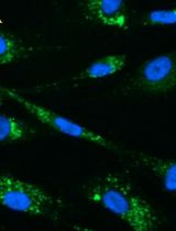
Accumulating evidence is revealing the essential role of immune system in cancer treatment. Certain chemotherapeutic drugs can potently induce the release of ‘cell death associated molecular patterns’ (CDAMPs), which accompanies cancer cell demise. CDAMPs can engage corresponding receptors on immune cells and stimulate immune responses to achieve long-term tumor control (Ma et al., 2013; Ma et al., 2014; Yang et al., 2015). Among reported CDAMPs, calreticulin (CALR), ATP and HMGB1 are well known for their immune-stimulatory effect. Here we describe the assays that we applied to measure cell death and these CDAMPs. Briefly, cell death can be analyzed by co-staining of 4’,6-diamidino-2-phenylindole (DAPI) with 3,3’-Dihexyloxacarbocyanine Iodide [DiOC6(3)] or Annexin V. CALR exposure on the cell membrane can be detected by flow cytometry. ATP and HMGB1 release can be quantified by luminescence assay and ELISA assay respectively.
Keywords: Cell death (细胞死亡)Background
Lactate dehydrogenase assay and trypan blue staining are traditional methods to examine cell death. We describe here two feasible and economic solutions to detect apoptotic and necrotic cell death by flow cytometry (FCM). DAPI labels cells with disrupted integrity (necrosis), while Annexin V binds to phosphatidylserine (which is externalized upon apoptosis). DiOC6(3) uptake indicates mitochondrial transmembrane potential (MTP) and the collapse of MTP reveals apoptosis. DAPI doesn’t require compensation with phycoerythrin (PE, which is conjugated with Annexin V protein) or DiOC6(3), and therefore show advantage over propidium iodide (PI) in these assays.
CALR is an endoplasmic reticulum protein, and activation of caspase cascades triggers CALR translocation to cytoplasmic membrane. CALR exposure can be detected by immunofluorescent staining with corresponding antibodies, followed by FCM- or microscopy-based detection. Extracellular ATP can be measured by enzymatic activity of Luciferase, while HMGB1 concentration can be detected by sensitive ELISA kits.
Materials and Reagents
- 24 well plates
- 2 ml Eppendorf tubes
- MCA205 fibrosarcoma cells (H2b) were induced by 3-methylcholanthrene in C57BL/6 mice (Shu et al., 1985). The assays described here are applicable for other cell lines
- DMEM high glucose (Thermo Fisher Scientific, GibcoTM, catalog number: 11965092 , or GE Healthcare, HyCloneTM , catalog number: SH30022 )
- Fetal bovine serum (FBS) (Thermo Fisher Scientific, GibcoTM, catalog number: 16000044 )
- 100 mM sodium pyruvate solution (Thermo Fisher Scientific, GibcoTM, catalog number: 11360070 )
- HEPES, 1 M buffer solution (Thermo Fisher Scientific, GibcoTM, catalog number: 15630080 )
- Penicillin and streptomycin
- Mitoxantrone (Sigma-Aldrich, catalog number: M6545 )
- Trypsin (0.25%) (Thermo Fisher Scientific, GibcoTM, catalog number: 15050065 )
- 1x PBS, pH 7.4 (Thermo Fisher Scientific, GibcoTM, catalog number: 10010031 )
- DiOC6(3) (3,3’-dihexyloxacarbocyanine Iodide) (Thermo Fisher Scientific, Molecular ProbesTM, catalog number: D273 )
- DAPI (4’,6-diamidino-2-phenylindole, dihydrochloride) (Thermo Fisher Scientific, Molecular ProbesTM, catalog number: D1306 )
- CALR antibody (Ab): rabbit monoclonal Ab, clone number: EPR3924 (Abgent, catalog number: AJ1124a ; or Abcam, catalog number: ab92516 ); or rabbit polyclonal Ab (Abcam, catalog number: ab2907 )
- Alexa488 conjugated goat anti-rabbit IgG (H+L) secondary antibody (Thermo Fisher Scientific, Invitrogen, catalog number: A-11008 )
- ATP Bioluminescent Assay Kit (Sigma-Aldrich, catalog number: FL-AA ) or ENLITEN® ATP Assay System (Promega, catalog number: FF2000 )
- Annexin V Apoptosis Detection Kit (BD, catalog number: 559763 )
- HMGB1 ELISA Kit (Tecan Trading, catalog number: ST51011 )
- 1x Annexin V binding buffer (see Recipes)
- ATP assay mix (see Recipes)
- rLuciferase/Luciferin buffer(see Recipes)
Equipment
- Centrifuge
- Water-Jacketed CO2 incubator (Thermo Fisher Scientific, Thermo ScientificTM, model: HERAcell® 150i )
- Multi-channel pipette
- SpectraMax L microplate reader (Molecular Device, model: SpectraMax L )
- SpectraMax i3 multi-mode detection platform (Molecular Device, model: SpectraMax i3 )
- AttuneTM NxT flow cytometer (Thermo Fisher Scientific, model: AttuneTM NxT Flow Cytometer )
Note: Alternatively, VICTORTM X multilabel reader (PerkinElmer) and MACSQuant® Analyzer 10 (Miltenyi Biotec) are also suitable.
Software
- Excel (Microsoft Office)
- GraphPad Prism (San Diego, CA, USA)
- FlowJo software (Treestar Inc., Ashland, OR, USA)
Procedure
文章信息
版权信息
© 2016 The Authors; exclusive licensee Bio-protocol LLC.
如何引用
Ma, Y. and Yang, H. (2016). In vitro Assays for the Detection of Calreticulin Exposure, ATP and HMGB1 Release upon Cell Death. Bio-protocol 6(24): e2076. DOI: 10.21769/BioProtoc.2076.
分类
癌症生物学 > 细胞死亡 > 免疫学试验
生物化学 > 其它化合物 > 三磷酸核苷
细胞生物学 > 细胞活力 > 细胞死亡
您对这篇实验方法有问题吗?
在此处发布您的问题,我们将邀请本文作者来回答。同时,我们会将您的问题发布到Bio-protocol Exchange,以便寻求社区成员的帮助。