

Analysis of Myosin II Minifilament Orientation at Epithelial Zonula Adherens
上皮粘着小带的微球蛋白II微丝排列分析


发布: 2016年12月05日第6卷第23期 DOI: 10.21769/BioProtoc.2054 浏览次数: 9674
评审: Nicoletta CordaniThomas J. BartoshXuecai Ge
参见作者原研究论文
The authors used this protocol in:
Apr 2016


Abstract
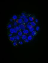
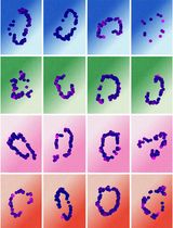
Non-muscle myosin II (NMII) form bipolar filaments, which bind F-actin to exert cellular contractility during physiological processes (Vicente-Manzanares et al., 2009). Using a combinatorial approach to fluorescently label both N- and C-termini of the NMII heavy chain, recent works have demonstrated the ability to visualize NMII bipolar filaments at various subcellular localizations (Ebrahim et al., 2013; Beach et al., 2014). At the zonula adherens (ZA) of epithelia, NMII minifilaments bind the circumferential actin bundles in a pseudo-sarcomeric manner (Ebrahim et al., 2013), a conformation required to maintain junctional tension and tissue integrity (Ratheesh et al., 2012). By expressing green fluorescent protein (GFP)-NMIIA heavy chain and immunolabel it using a NMIIA C-terminus specific antibody, we were able to visualize the NMII minifilaments bound to F-actin bundles in Caco-2 cells (Michael et al., 2016), as previously reported (Ebrahim et al., 2013; Beach et al., 2014). In addition, we designed an FIJI/MATLAB analysis module to quantify the size, distance and alignment of these minifilaments with respect to junctional F-actin at the ZA. Measurements of the dispersion of minifilaments angles were proven to be a useful parameter that closely correlated to the extent of contractility at junctions (Michael et al., 2016).
Keywords: Myosin II minifilaments (肌球蛋白II微丝)Background
For decades, the assembly of NMII into bipolar filaments has been studied using electron microscopy (EM) techniques. These mainly involve the assembly of NMII minifilaments from purified proteins or the visualization of minifilaments in cells following extraction of the actin cytoskeleton (Pollard, 1982; Svitkina et al., 1989). Whilst these methods enabled measurements of NMII bipolar filament assemblies, they were technically challenging and did not accurately reflect the cellular distribution of these entities, notwithstanding the artifacts introduced due to the sample preparation. With the advent of super-resolution microscopy, we are now able to observe and measure these NMII minifilaments in various subcellular locations with high resolution, using a rapid process that is amenable for most laboratories equipped with a microscope that performs structured illumination microscopy (SIM, Yap et al., 2015). In this protocol, we describe a method that we have developed to assess NMII minifilaments properties at adherens junctions by measuring the lengths of the minifilaments as well as quantifying their angles with respect to the junctional F-actin and their distance from the junctions (Michael et al., 2016).
Materials and Reagents
- 1.6-well plates (Corning, Costar®, catalog number: 3516 )
- Glass coverslips (13 mm, #1.5) (Thermo Fisher Scientific, Thermo ScientificTM, catalog number: 1014355130NR15 )
- ShandonTM ColorFrostTM glass slides (Thermo Fisher Scientific, Thermo ScientificTM, catalog number: 6776214 )
- Caco-2 human colon adenocarcinoma cells (ATCC, catalog number: ATCCH®TB-37TM )
- Plasmid: GFP-NMIIA (Addgene, catalog number: 11347 )
- Roswell Park Memorial Institute (RPMI) 1640 medium (Thermo Fisher Scientific, GibcoTM, catalog number: 11875093 )
- Fetal bovine serum (FBS) (Thermo Fisher Scientific, GibcoTM, catalog number: 26140079 )
- 100x penicillin/streptomycin (10,000 U/ml) (Thermo Fisher Scientific, GibcoTM, catalog number: 15140122 )
- 100x L-glutamine (200 mM) (Thermo Fisher Scientific, GibcoTM, catalog number: 25030081 )
- Lipofectamine® 3000 (Thermo Fisher Scientific, InvitrogenTM, catalog number: L3000015 )
- Opti-MEM®, reduced serum media (Thermo Fisher Scientific, GibcoTM, catalog number: 31985070 )
- Alexa Fluor® 647 phalloidin (Thermo Fisher Scientific, Molecular ProbesTM, catalog number: A22287 )
- Triton X-100 (Sigma-Aldrich, catalog number: X100 )
- Antibodies
- Anti-Myosin IIA rabbit polyclonal antibody (Biolegend, catalog number: PRB-440P )
Note: This antibody targets the C-terminal portion of the NMIIA heavy chain. - Goat anti-Rabbit IgG (H+L) secondary antibody, Alexa Fluor® 546 conjugate (Thermo Fisher Scientific, Invitrogen, catalog number: A11035 )
- TetraSpeckTM multi-speck 100 nm beads (Thermo Fisher Scientific, Molecular ProbesTM, catalog number: T7279 )
- ProLong® Gold antifade mountant (Thermo Fisher Scientific, Molecular ProbesTM, catalog number: P36934 )
- Paraformaldehyde (PFA) (Sigma-Aldrich, catalog number: 158127 )
- Piperazine-N,N’-bis(2-ethanesulfonic acid) (PIPES) (Sigma-Aldrich, catalog number: P6757 )
- Potassium chloride (KCl) (Sigma-Aldrich, catalog number: P9541 )
- Sucrose (Sigma-Aldrich, catalog number: S0389 )
- Ethylene glycol-bis(β-aminoethyl ether)-N,N,N’,N’-tetraacetic acid (EGTA) (Sigma-Aldrich, catalog number: E3889 )
- Magnesium chloride (MgCl2) (Sigma-Aldrich, catalog number: M8266 )
- Tris-Cl (Sigma-Aldrich, catalog number: T5941 )
- Sodium chloride (NaCl) (Sigma-Aldrich, catalog number: S3014 )
- Bovine serum albumin (BSA) (Sigma-Aldrich, catalog number: A2153 )
- Phosphate buffered saline (PBS), without Ca2+ and Mg2+ (Thermo Fisher Scientific, GibcoTM, catalog number: 14190250 )
- 10x trypsin/EDTA (0.5%) (Thermo Fisher Scientific, GibcoTM, catalog number: 15400054 )
- 4% paraformaldehyde (PFA) (see Recipes)
- Tris buffered saline (TBS) (see Recipes)
- Blocking buffer (see Recipes)
Equipment
- NuncTM 75 cm2 cell culture flasks (Thermo Fisher Scientific, Thermo ScientificTM, catalog number: 156472 )
- Incubator
- Zeiss ELYRA superresolution microscope (ZEISS, model: ELYRA Superresolution Microscope )
Software
- Zen (black version; Zeiss)
- FIJI (http://imagej.net/Fiji)
- Prism, GraphPad (http://www.graphpad.com/scientific-software/prism/)
- Matlab, Mathworks (https://www.mathworks.com/index-c.html)
- FIJI and Matlab scripts for minifilament analysis (see Appendix I and II of this Bioprotocol)
Procedure
文章信息
版权信息
© 2016 The Authors; exclusive licensee Bio-protocol LLC.
如何引用
Michael, M., Liang, X. and Gomez, G. A. (2016). Analysis of Myosin II Minifilament Orientation at Epithelial Zonula Adherens. Bio-protocol 6(23): e2054. DOI: 10.21769/BioProtoc.2054.
分类
细胞生物学 > 细胞结构 > 细胞粘附
细胞生物学 > 细胞成像 > 荧光
您对这篇实验方法有问题吗?
在此处发布您的问题,我们将邀请本文作者来回答。同时,我们会将您的问题发布到Bio-protocol Exchange,以便寻求社区成员的帮助。