- EN - English
- CN - 中文
Resin-embedded Thin-section Immunohistochemistry Coupled with Triple Cellular Counterstaining
树脂包埋薄层免疫组织化学法联合三重细胞复染
发布: 2017年04月05日第7卷第7期 DOI: 10.21769/BioProtoc.2052 浏览次数: 8220
评审: Tie LiuGaston A. PizzioFang Xu
Abstract
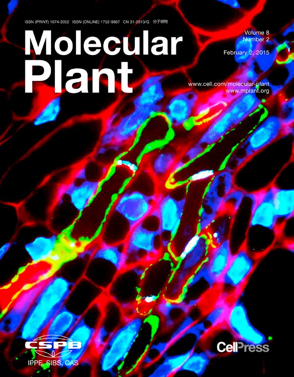
This protocol was developed to study protein localisation within the vascular bundles of developing tomato fruit however, it can be applied to any resin embedded plant tissue. The vascular bundle is comprised of many different cells that all have unique properties. The mature sieve elements are enucleated and contain sieve plates that comprise of callose. This method has utilised these properties of the sieve element by combining immunohistochemistry for cell wall invertase with counterstaining of aniline blue for callose, DAPI for nucleus and cell structure is shown with the final staining of the cell wall using calcofluor white. It must be noted that when following this protocol, it is vital for the sections to be flat and fixed to the slide with gelatine so cover slip removal does not move the sample section. This protocol will be applicable to all plant tissues and provides additional evidence of the protein localisation within the cell by conducting a counterstaining procedure.
Keywords: Immunolocalisation (免疫定位)Background
Immunolocalisation has long been a method used to study the localisation of proteins within tissue. This protocol focused on, not only localising the proteins of interest but also the molecular structures that were in surrounding tissue. It is believed that the mature sieve elements are enucleated and have abundant callose deposition within the sieve plate. Therefore, counterstaining procedures were applied in order to represent these biological phenomena. As the proteins of interest in this study were also thought to be localised within the apoplast further counterstaining was applied showing co-labelling of the protein and the cell wall.
Materials and Reagents
- Size 000 gelatine capsules (ProSciTech, catalog number: RL039 )
- Razor blade
- Microscope slide (Livingstone, catalog number: 7107-PPN )
- 22 x 50 mm, 0.17 mm thick coverslip (Fisher Scientific, catalog number: 12-543C )
- Tomato flowers 2 Days Before Fertilization (DBF) and 2 Days After Fertilization (DAF) harvested from glasshouse-grown cv. Moneymaker tomato plants
- 50 mM PIPES (Sigma-Aldrich, catalog number: F6757 )
- AR grade EtOH (Sigma-Aldrich, catalog number: 32205 )
Note: This product has been discontinued. - ddH2O
- LR white resin (ProSciTech, catalog number: C023 )
- Gelatine (Sigma-Aldrich, catalog number: G9391 )
- Cell wall invertase (LIN5) purified polyclonal primary antibody – produced in rabbit (Mimmitopes – custom made)
- Inhibitor of invertase (INH) purified polyclonal primary antibody – produced in rabbit (Mimmitopes – custom made)
- TBST (Sigma-Aldrich, catalog number: T9039 )
- Secondary anti-rabbit IgG fluorescein isothiocyanate (FITC) (Sigma-Aldrich, catalog number: F9887 )
- Anti-Rabbit IgG (whole molecule)-FITC antibody produced in goat (Sigma-Aldrich, catalog number: F6005 )
Note: This product has been discontinued. - Aniline blue (0.1% in ddH2O) (Sigma-Aldrich, catalog number: B8563 )
- Mowiol-phenylenediamine (mowiol) (Sigma-Aldrich, catalog number: 10852 )
- 4’,6-diamidino-2-phenylindole (DAPI) (1:500) (Sigma-Aldrich, catalog number: D9542 )
- Calcofluor white (0.1% in ddH2O) (Sigma-Aldrich, catalog number: F3543 )
- 2% paraformaldehyde
- Glutaraldehyde
- CaCl2
- Tris
- NaN3
- Bovine serum albumin (BSA)
- Fixing solution (see Recipes)
- Blocking buffer (see Recipes)
Equipment
- Rotator
- Dissecting microscope or magnifying glass
- Reichert Ultracut E microtome (Reichert, model: 701704 )
- DiATOME Histo Knife, Diamond, 45°, 4.0-4.9 mm (ProSciTech, catalog number: UH45-40 )
- Fume hood
- Beaker
- Axio Scope.A1 epifluorescence compound microscope (ZEISS, model: Axio Scope.A1 )
- Emission FITC filter (50-490 nm excitation, long pass 515 nm)
- Emission UV filter (365 nm excitation, short pass 420 nm)
- AxioCam digital camera (ZEISSTM) or equivalent
Software
- AxioVision V4.8 software
- Adobe Bridge CS4 software
- Abode Photoshop CS4 software
Procedure
文章信息
版权信息
© 2017 The Authors; exclusive licensee Bio-protocol LLC.
如何引用
Palmer, W. M., Patrick, J. W. and Ruan, Y. (2017). Resin-embedded Thin-section Immunohistochemistry Coupled with Triple Cellular Counterstaining. Bio-protocol 7(7): e2052. DOI: 10.21769/BioProtoc.2052.
分类
植物科学 > 植物细胞生物学 > 细胞成像
细胞生物学 > 细胞成像 > 固定组织成像
您对这篇实验方法有问题吗?
在此处发布您的问题,我们将邀请本文作者来回答。同时,我们会将您的问题发布到Bio-protocol Exchange,以便寻求社区成员的帮助。
Share
Bluesky
X
Copy link


