

Vascular Smooth Muscle Cell Isolation and Culture from Mouse Aorta
小鼠主动脉血管平滑肌细胞的分离和培养
发布: 2016年12月05日第6卷第23期 DOI: 10.21769/BioProtoc.2045 浏览次数: 23424
评审: Jia LiGuillermo GomezAnonymous reviewer(s)
参见作者原研究论文
The authors used this protocol in:
Mar 2016


Abstract
Vascular smooth muscle cells (SMC) in the ascending thoracic aorta arise from neural crest cells, whereas SMCs in the descending aorta are derived from the presomitic mesoderm. SMCs play important roles in cardiovascular development and aortic aneurysm formation. This protocol describes the detailed process for explanting ascending and descending SMCs from mouse aortic tissue. Conditions for maintenance and subculture of isolated SMCs and characterization of the vascular SMC phenotype are also described.
Keywords: Tissue culture (组织培养)Background
Vascular smooth muscle cells (SMCs) make up the muscular medial layer of arteries. Larger elastic arteries, such as the aorta, have multiple concentric lamellae consisting of aligned smooth muscle cells sandwiched between elastin fibers. The elastin and collagen present within the medial layer of elastic arteries allow it to distribute the force generated by the heart throughout the vessel wall (Wagenseil and Mecham, 2009). Smaller muscular arteries, by contrast, have only an internal and external elastic lamina bounding the smooth muscle layer. These arteries are downstream in the arterial tree and thus bear less force from blood flow.
Vascular smooth muscle cells, unlike cardiac and skeletal muscle cells, are capable of modulating their phenotype in response to vascular injury or environmental cues. Under normal physiologic conditions, quiescent, contractile SMCs populate the artery wall and contract to regulate vascular tone and keep blood flow continuous in response to pulsatile pressures. Contractile SMCs are characterized by high expression of smooth muscle-specific contractile genes, including smooth muscle specific α-actin and myosin heavy chain (Owens et al., 2004). However, in response to injury or mitogenic stimuli, SMCs downregulate expression of the contractile genes and take on a synthetic phenotype: they proliferate rapidly, migrate into the site of injury, and remodel the extracellular matrix by synthesizing both matrix-digesting enzymes and new matrix proteins. Many vascular diseases are associated with a synthetic SMC phenotype, including atherosclerosis (Owens, 1995).
SMCs located in different areas of the body actually arise from diverse embryonic lineages (Majesky, 2007). For example, the SMCs populating the ascending thoracic aorta and the cerebrovasculature are derived from neural crest cells. However, SMCs in the descending thoracic aorta come from mesodermal origins. These distinctions affect the ultimate properties of the SMCs, so it is important when designing experiments to use SMCs from the same developmental origin where the phenotype of interest arises.
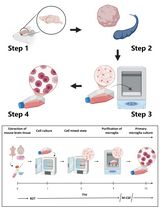
In this protocol, we give detailed instructions for isolating and culturing SMCs from the ascending and descending thoracic aortas in the mouse. We have previously used this technique to isolate SMCs from genetically modified mice and their wild-type littermates to provide an in vitro system for looking at the effect of genetic changes on SMC phenotype (Cao et al., 2010; Kuang et al., 2012; Kuang et al., 2016; Papke et al., 2013; Kwartler et al., 2014).
Materials and Reagents
- For initial explant
- Dissection material: 2 sterile forceps, 2 sterile tweezers, 1 sterile scissor, 2 sterile scalpels
- Disposable scalpels (Aspen Surgical, catalog number: 371621 )
- Four 60 mm tissue culture dishes per sample (Corning, catalog number: 430589 )
- Syringe, 10 ml
- Syringe filter, 0.22 µm pore
- 500 ml filtration unit, 0.22 µm pore (EMD Millipore, catalog number: SCGPU01RE )
- Parafilm
- Aluminum foil
- 2.5% avertin-2,2,2-tribromoethanol (Sigma-Aldrich, catalog number: T48402-25g ) arrives as powder, make 100% stock solution by dissolved 25 g 2,2,2-tribromoethanol in 25 ml of 2-methyl-2-butanol (Sigma-Aldrich, catalog number: 240486-100ML ), then dilute to 2.5% solution in sterile water. Both the stock solution and the dilution are light sensitive, so should be stored in the dark at 4 °C
- 70% ethanol
- Dulbecco’s phosphate buffered saline (DPBS) with calcium and magnesium (GE Healthcare, HycloneTM, catalog number: SH30028.02 )
- Components of aorta biopsy storage media:
- Waymouth’s MB 752/1 medium, 500 ml (Thermo Fisher Scientific, GibcoTM, catalog number: 11220035 )
- Antibiotic-antimycotic, 100x (Sigma-Aldrich, catalog number: A5955 )
- L-glutamine, 100x (Sigma-Aldrich, catalog number: G7513 )
- Sodium bicarbonate (Sigma-Aldrich, catalog number: S8761 )
- MEM non-essential amino acids (Sigma-Aldrich, catalog number: M7145 )
- HEPES buffer (Sigma-Aldrich, catalog number: H0887 )
- Collagenase type 1 (Sigma-Aldrich, catalog number: C1639 )
- Elastase, Pancreatic type 1 from porcine pancreas (Sigma-Aldrich, catalog number: E1250 )
- Soybean trypsin inhibitor (Thermo Fisher Scientific, GibcoTM, catalog number: 17075-029 )
- Heat inactivated fetal bovine serum (FBS) (Atlanta Biologicals)
- SmBm bullet kit (Lonza, catalog number: CC3182 )
- FGF
- EGF
- Insulin
- Gentamicin and included FBS - Do not use!
- For continued culture
- 500 ml filtration unit, 0.22 µm pore (EMD Millipore, catalog number: SCGPU01RE )
- Freeze vials (2 ml)
- Fetal bovine serum (FBS, Atlanta Biologicals)
- Antibiotic-antimycotic, 100x (Sigma-Aldrich, catalog number: A5955 )
- SmBm bullet kit (Lonza, catalog number: CC3182 )
- FGF
- EGF
- Insulin
- FGF
- TrypLE express (Thermo Fisher Scientific, GibcoTM, catalog number: 12604013 )
- DMSO
- For immunofluorescence to confirm SMC identity
- Coverslips (UV treated, please see Data analysis section below for details)
- 6 well plates
- Hemocytometer
- 3-6 mice, preferably aged 4-6 weeks old
- SmBm basal media (Lonza, catalog number: CC3181 )
- Fetal bovine serum (FBS) (Atlanta Biologicals)
- Recombinant human TGF-β1 (rhTGF- β1, R&D Systems)
- 16% formaldehyde (Thermo Fisher Scientific, Thermo ScientificTM, catalog number: 28906 ). Dilute 1 to 4 to a final concentration of 4% in DPBS with calcium and magnesium
- Blocking buffer (0.3% Triton X, 0.5% BSA in DPBS)
- DPBS
- Smooth muscle α-actin antibody (Sigma-Aldrich, catalog number: A5228 )
- Anti-mouse secondary antibody conjugated with fluorescence
- Mounting medium with DAPI (Vectashield, catalog number: H-1200 )
- Aorta biopsy storage media (see Recipes)
- Complete smooth muscle media (see Recipes)
- Smooth muscle cell freeze media (see Recipes)
- Digestive enzyme mix (see Recipes)
Equipment
Note: The reproducibility of the experiment is not dependent on the specific brand or model of these equipment. Any microscope, tissue culture hood/incubator, centrifuge, etc., will give similar results.
- Magnetic stirrer
- Dissecting microscope
- Sterile tissue culture hood
- Sterile tissue culture incubator
- 10 ml pipet
- Water bath
- Tabletop centrifuge
- 200 µl pipette
- T25 flasks
- T75 flasks
- Slow freeze container (either a foam container or an isopropanol based container will work)
- Hemocytometer
- Inverted microscope
- Hot bead sterilizer
Procedure
文章信息
版权信息
© 2016 The Authors; exclusive licensee Bio-protocol LLC.
如何引用
Kwartler, C. S., Zhou, P., Kuang, S., Duan, X., Gong, L. and Milewicz, D. M. (2016). Vascular Smooth Muscle Cell Isolation and Culture from Mouse Aorta. Bio-protocol 6(23): e2045. DOI: 10.21769/BioProtoc.2045.
分类
发育生物学 > 细胞生长和命运决定 > 血管生成
细胞生物学 > 细胞分离和培养 > 细胞分离
细胞生物学 > 细胞分离和培养 > 细胞分化
您对这篇实验方法有问题吗?
在此处发布您的问题,我们将邀请本文作者来回答。同时,我们会将您的问题发布到Bio-protocol Exchange,以便寻求社区成员的帮助。