

Preparation of Purified Gram-positive Bacterial Cell Wall and Detection in Placenta and Fetal Tissues
纯化制备及检测胎盘和胎儿组织中革兰氏阳性细菌的细胞壁


发布: 2016年12月05日第6卷第23期 DOI: 10.21769/BioProtoc.2037 浏览次数: 10824
评审: Alexander B. WestbyeMigla MiskinyteAnonymous reviewer(s)
参见作者原研究论文
The authors used this protocol in:
Mar 2016
Advertisement


Abstract
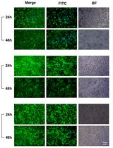
Cell wall is a complex biopolymer on the surface of all Gram-positive bacteria. During infection, cell wall is recognized by the innate immune receptor Toll-like receptor 2 causing intense inflammation and tissue damage. In animal models, cell wall traffics from the blood stream to many organs in the body, including brain, heart, placenta and fetus. This protocol describes how to prepare purified cell wall from Streptococcus pneumoniae, detect its distribution in animal tissues, and study the tissue response using the placenta and fetal brain as examples.
Keywords: Cell wall (细胞壁)Background
Host response to infection involves recognition of many bacterial components including the cell wall (CW), a complex macromolecule that forms the surface of all Gram-positive bacteria. The CW of Gram-positive bacteria is formed by the covalent network of peptidoglycan and teichoic acid. Streptococcus pneumoniae, a leading cause of pneumonia, sepsis, and meningitis, has served as an important model organism for studying the innate immune response to Gram-positive bacterial infection including CW.
When upon Streptococcus pneumoniae (pneumococcal) infection CW components are released from bacteria during growth or antibiotic-induced death, it circulates in the blood stream and crosses cellular barriers, including the placenta and blood brain barrier. CW components have inflammatory activities equal to or greater than intact bacteria (Tuomanen et al., 1985a and 1985b). The CW can be viewed as the Gram-positive equivalent of endotoxin. The vast amount of CW pieces released during infection greatly stimulates the host inflammatory response by activating the innate immune receptor, Toll like receptor 2 (TLR2) (Yoshimura et al., 1999). Responses differ depending on the organ infected: the postnatal brain undergoes apoptosis, scarring predominates in heart, and the fetal brain escapes damage, showing striking neuroproliferation (Orihuela et al., 2006; Braun et al., 1999; Fillon et al., 2006; Humann et al., 2016).
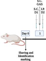
This protocol describes how to prepare purified CW from Streptococcus pneumoniae (Tuomanen et al., 1985b) and follow its distribution in mice after intravenous injection, focusing on the placenta and fetal brain as examples (Humann et al., 2016). This model yields histopathologic sections of organs for study of the tissue response to CW components. Our model’s focus on pneumococcal CW derives from its well-described role in inflammation and injury in many organs, its extensive known chemistry and its recognition as a classic TLR2 pathogen associated molecular pattern.
Materials and Reagents
- 0.22 µm bottle top filter (Corning, catalog number: 431096 )
- Glass tubes (Thermo Fisher Scientific, Fisher Scientific, catalog number: 14-961-32 )
- 1,000 ml centrifuge bottle (Thermo Fisher Scientific, Thermo ScientificTM, catalog number: 3120-1000 )
- 30 ml centrifuge bottle (Thermo Fisher Scientific, Thermo ScientificTM, catalog number: 3119-0050 )
- Serological pipettes, 1 case of 5 ml, 10 ml, 25 ml
- Microcentrifuge tubes (Eppendorf, catalog number: 022364111 )
- Aluminum foil (Thermo Fisher Scientific, Fisher Scientific, catalog number: 01-213-100 )
- 25 gauge needles (BD, catalog number: 305122 )
- 50 ml polypropylene tubes (SARSTEDT, catalog number: 62.547.254 )
- Petri dishes (Thermo Fisher Scientific, Fisher Scientific, catalog number: FB0875712 )
- Small electric razor to shave animals
- Superfrost microslides (VWR, catalog number: 48311-703 )
- Premium cover glass (Thermo Fisher Scientific, Fisher Scientific, catalog number: 12-548-5P )
- Magnetic stir bars
- Inoculation loops (Thermo Fisher Scientific, Fisher Scientific, catalog number: 22-363-595 )
- 1 ml syringe (BD, catalog number: 309628 )
- Acid washed 106 µm glass beads (Sigma-Aldrich, catalog number: G4649 )
- Plastic embedding molds for histology (Polysciences, catalog number: 18646D-1 )
- Streptococcus pneumoniae strains - CW is much easier to purify from strains that are unencapsulated such as R6 (ATCC, catalog number: BAA-255 )
- C57Bl6 mice, mixture of male and female (THE JACKSON LABORATORY, catalog number: 000664 )
- Water (Sigma-Aldrich, catalog number: W3500-1L )
- Ultrapure water (example: Milli-Q, EMD Millipore)
- Tryptic soy agar (TSA) (EMD Millipore, catalog number: 105458 )
- Sterile defibrinated sheep blood (i-Tek Medical Technologies, catalog number: 103-100-3 )
- Glycerol
- Sodium dodecyl sulfate (SDS) (Sigma-Aldrich, catalog number: L4390 )
Note: This product has been discontinued. - Sodium chloride (NaCl) (Sigma-Aldrich, catalog number: 71382 )
- Magnesium sulfate (MgSO4) (Sigma-Aldrich, catalog number: 208094 )
- DNase I (Sigma-Aldrich, catalog number: DN25-10MG )
- RNase A (Sigma-Aldrich, catalog number: R6513-10MG )
- Calcium chloride dihydrate (CaCl2·2H2O) (Sigma-Aldrich, catalog number: C-3881 )
- Trypsin (AMRESCO, catalog number: M150 )
- α-amylase (Sigma-Aldrich, catalog number: A3176 )
- Lithium chloride (LiCl) (Sigma-Aldrich, catalog number: L9650 )
- Ethylenediaminetetraacetic acid disodium salt dehydrate (EDTA) (Sigma-Aldrich, catalog number: E5134 )
- Acetone (Thermo Fisher Scientific, Fisher Scientific, catalog number: A946-4 )
- PierceTM LAL Chromogenic Endotoxin Quantitation Kit (Thermo Fisher Scientific, Thermo ScientificTM, catalog number: 88282 )
- Fluorescein isothiocyanate isomer I (FITC) (Sigma-Aldrich, catalog number: F7250 )
- Dulbecco’s phosphate buffered saline (DPBS) (Mediatech, catalog number: 21-030- CV )
- Paraformaldehyde, 16% w/v (Alfa Aesar, catalog number: 43368 )
- Sucrose (Sigma-Aldrich, catalog number: S9378 )
- Tissue Tek OCT compound (SAKURA FINETEK USA, catalog number: 4583 )
- Prolong Gold Antifade with DAPI (Thermo Fisher Scientific, Molecular ProbesTM, catalog number: P36931 )
- Trizma HCl (Sigma-Aldrich, catalog number: T5941 )
- Tris-base (Sigma-Aldrich, catalog number: 1070897600 )
- Hydrochloric acid (HCl) (Sigma-Aldrich, catalog number: 320331-500ml )
- Sodium carbonate (Na2CO3) (Sigma-Aldrich, catalog number: 451614 )
- Sodium bicarbonate (NaHCO3) (Sigma-Aldrich, catalog number: S-6014 )
- Magnesium chloride hexahydrate (MgCl2) (Sigma-Aldrich, catalog number: M0250 )
- Calcium chloride (CaCl2) (Sigma-Aldrich, catalog number: C1016 )
- MnSO4 monohydrate (Sigma-Aldrich, catalog number: M7634 )
- Glucose (Sigma-Aldrich, catalog number: G7528 )
- Adenosine (Oakwood Products, catalog number: 093333 )
- Uridine (EMD Millipore, catalog number: 6680 )
- Glutamine (Sigma-Aldrich, catalog number: G8540 )
- Nicotinic acid (Sigma-Aldrich, catalog number: N4126 )
- (B6) Pyridoxine HCl (Sigma-Aldrich, catalog number: P9755 )
- Ca-pantothenate (D-pantothenic acid) (Sigma-Aldrich, catalog number: P3161 )
Note: This product has been discontinued. - Thiamine HCl (Sigma-Aldrich, catalog number: 5871-100GM )
- Riboflavin (Sigma-Aldrich, catalog number: R4500 )
- Biotin (Sigma-Aldrich, catalog number: B4639 )
- 10 N NaOH solution (Thermo Fisher Scientific, Fisher Scientific, catalog number: SS255-1 )
- Ferrous sulfate heptahydrate (FeSO4·7H2O) (Thermo Fisher Scientific, Fisher Scientific, catalog number: I146-500 )
- Copper(II) sulfate pentahydrate (CuSO4·5H2O) (Sigma-Aldrich, catalog number: C8027 )
- Zinc sulfate heptahydrate (ZnSO4·7H2O) (Sigma-Aldrich, catalog number: Z4750 )
- Manganese chloride tetrahydrate (MnCl2·4H2O) (Thermo Fisher Scientific, Fisher Scientific, catalog number: M87-500 )
- Pyruvic acid (Sigma-Aldrich, catalog number: 107360-25g )
- Potassium phosphate monobasic (KH2PO4) (Sigma-Aldrich, catalog number: P5655 )
- Potassium phosphate dibasic (K2HPO4) (Sigma-Aldrich, catalog number: P3786 )
- Yeast extract (BD, BactoTM, catalog number: 212750 )
- Sodium acetate anhydrous (Sigma-Aldrich, catalog number: S8625 )
- Casamino acids technical (BD, BactoTM, catalog number: 223120 )
- L-tryptophan (Sigma-Aldrich, catalog number: T8941 )
- L-cysteine HCl (Thermo Fisher Scientific, Fisher Scientific, catalog number: BP376-100 )
- Asparagine (Sigma-Aldrich, catalog number: A4284 )
- Choline chloride (Sigma-Aldrich, catalog number: C7527 )
- Dry ice
- Solutions used to treat CW during purification
- 50 mM Tris-HCl, pH 7.0 (see Recipes)
- 5% (v/v) SDS (see Recipes)
- 1 M NaCl (see Recipes)
- 100 mM Tris, pH 7.5 (see Recipes)
- 1 M MgSO4 (see Recipes)
- 1 M CaCl2 (see Recipes)
- 1% (v/v) SDS (see Recipes)
- 8 M LiCl (see Recipes)
- 100 mM EDTA (see Recipes)
- Carbonate buffer, pH 9.2 (see Recipes)
- Preparation of C+Y components
- ‘3 in 1’ salts (see Recipes)
- 20% glucose (see Recipes)
- 50% sucrose (see Recipes)
- Adenosine (2 mg/ml) (see Recipes)
- Uridine (2 mg/ml) (see Recipes)
- Glutamine (1 mg/ml) (see Recipes)
- Adams I (see Recipes)
- Adams II (see Recipes)
- 2% pyruvate (see Recipes)
- 1 M KH2PO4 (see Recipes)
- 1 M K2HPO4 (see Recipes)
- 5% yeast extract (see Recipes)
- Media preparation
- PreC media (see Recipes)
- Supplement (see Recipes)
- Adams III (see Recipes)
- 1 M potassium phosphate buffer (see Recipes)
- C+Y medium (see Recipes)
Equipment
- 37 °C CO2 incubator (Thermo Fisher Scientific, Thermo ScientificTM, model: 3110 )
- Benchtop microcentrifuge (Eppendorf, model: 5417C )
- 1,000 ml Erlenmeyer flasks
- 4,000 ml Erlenmeyer flasks
- Sorvall centrifuge RC 5C Plus and appropriate rotor for the centrifuge tubes or bottles
- 500 ml beaker
- Stirring hotplate
- Vortex mixer
- Speed-vac (Savant, model: SC110A )
- Water-bath sonicator (Thermo Fisher Scientific, Fisher Scientific, model: FS20 )
- Heating pad
- Cryostat microtome (Microme, model: HM505E )
- Zeiss LSM 510 NLO Meta confocal microscope
- Spectrophotometer (Turner, Model: 340 )
- Spectra MAX340 plate reader to measure absorbance at 405 nm (Molecular Device, model: Spectra MAX340 )
Software
- Zen 2008 software package (Carl Zeiss MicroImaging, Inc.)
- ImageJ (imagej.net/Particle_Analysis)
- Graphpad Prism (Graphpad)
Procedure
文章信息
版权信息
© 2016 The Authors; exclusive licensee Bio-protocol LLC.
如何引用
Mann, B., Loh, L. N., Gao, G. and Tuomanen, E. (2016). Preparation of Purified Gram-positive Bacterial Cell Wall and Detection in Placenta and Fetal Tissues. Bio-protocol 6(23): e2037. DOI: 10.21769/BioProtoc.2037.
分类
微生物学 > 体内实验模型 > 细菌
生物化学 > 脂质 > 脂质分离
您对这篇实验方法有问题吗?
在此处发布您的问题,我们将邀请本文作者来回答。同时,我们会将您的问题发布到Bio-protocol Exchange,以便寻求社区成员的帮助。