- EN - English
- CN - 中文
Fusarium graminearum Maize Stalk Infection Assay and Associated Microscopic Observation Protocol
玉米秸秆禾谷镰刀菌感染分析和显微镜观察
发布: 2016年12月05日第6卷第23期 DOI: 10.21769/BioProtoc.2034 浏览次数: 12367
评审: Arsalan DaudiBaohua LiKumiko Okazaki
Abstract
The ascomycete fungus Fusarium graminearum (previously also called Gibberella zeae) causes Gibberella stalk rot in maize (Zea mays) and results in lodging and serious yield reduction. To develop methods to assess the fungal growth and symptom development in maize stalks, we present here a protocol of maize stalk inoculation with conidiospores of fluorescent protein-tagged F. graminearumand microscopic observation of the stalk infection process. The inoculation protocol provides repeatable results in stalk rot symptom development, and allows tracking of fungal hyphal growth inside maize stalks at cellular scale.
Keywords: Fusarium graminearum (禾谷镰刀菌)Background
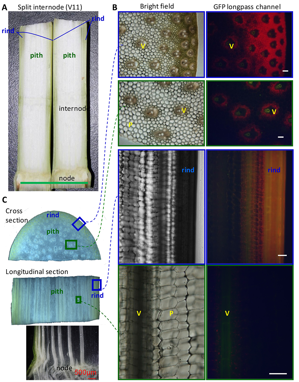
Maize (Zea mays) is one of the most important crops across the world. The stalk is the main stem of a maize plant. It is composed of nodes and internodes (Figure 1). The internodes comprise rind and pith, and vascular tissues are scattered in the pith. Figure 1 shows the microscopic images of maize stalk sections under bright field or GFP fluorescence channel. This provides context for maize stalk inoculation and observation.
The ascomycete fungus Fusarium graminearum (previously also called Gibberella zeae) causes Gibberella stalk rot in maize (Zea mays) and results in lodging and yield loss (Jackson et al., 2009; Santiago et al., 2007). F. graminearumcan also cause seedling blight and ear rot of maize, and Fusarium head blight of wheat and barley (Jackson et al., 2009). In the field, ascospores of F. graminearumoverwinter on infected crop residues such as maize stalks and wheat straw, and may infect other plants through wounds (e.g., caused by hail or pests), or may enter through young roots, and start a new infection cycle (Jackson et al., 2009). Although the entering routes and disease development time course may differ, the final symptom for maize Gibberella stalk rot is in the stalk of adult maize plant, which directly leads to lodging and yield loss. 
Figure 1. Anatomy of maize stalk. Uninfected internodes of maize stalk at V11 stage shown as reference for understanding F. graminearumprogression. V: vascular bundle. P: parenchyma cells. Green bar = 5 cm; White bars = 100 μm.
Compared to the inoculation method for assessing wheat head blight development (Pritsch et al., 2000 and 2001; Proctor et al., 1995), the inoculation method for maize stalk rot is less well established. To assess plant resistance or fungal virulence regarding maize stalk rot, two major types of inoculation methods have been reported. One is inoculation of young roots (Yang et al., 2010), which mimics the route of one type of natural infection, but has great variance in the time before stalk symptoms develop among individual maize plants, which makes follow-up microscopic observations difficult. The other is inoculation of mature stalk by wounding, usually at the internode immediately below the tassel (Zhou et al., 2010; Zheng et al., 2012) or at the internode immediately above aerial root node (Reid et al., 1996; Santiago et al., 2007) and then assessing lesions after 14 days. Recently we reported a method of wounding inoculation of maize stalks at lower internodes in combination with fluorescent protein-expressing fungi, which provided more synchronic symptom development for microscopic tracking of the infection process at the cellular scale (Zhang et al., 2016).
Materials and Reagents
- Sterile gauzes (regular cotton yarn 21s, 100% absorbent cotton, for medical use, many brands will work, we used 500 g pack from Shanghai HongLong Medical Material Company)
- Sterile 250 ml-centrifuge bottles (Beckman Coulter, catalog number: 356011 )
- 1.5 ml sterile centrifuge tube (Corning, Axygen®, catalog number: MCT-150-C )
- 500 ml flasks
- Single-side razor blades (CLOUD, YIZUN BRAND, catalog number: DD75 )
- Microscope slides (Grale Scientific, Sail Brand, catalog number: 7103 )
- Microscope cover slides (CITOTEST LABWARE MANUFACTURING, catalog number: 80330-2810 )
- Fungal strains: The F. graminearum strain PH-1 expressing fluorescent protein AmCyan under the promoter of VM3 from Neurospora crassa (AmCyanPH-1) (Yuan et al., 2008; Zhang et al., 2012).
- Plant material: Maize (Zea mays ssp. mays L.) cultivar B73 plants (Schnable et al., 2009) were cultivated in a phytotron at 22-26 °C with 65% relative humidity and a 14 h photoperiod for 8 weeks until the tenth leaf appeared.
- Glycerol (Sinopharm Chemical Reagent, catalog number: 10010618 )
- V8 vegetable juice (Campbell Soup Company, catalog number: V8® ORIGINAL )
- CaCO3
- 1.5% agar powder
- Mung bean
- V8 juice agar medium (see Recipes)
- Mung bean liquid medium (see Recipes)
Equipment
- Growth chamber (Jiangnan, model: RXZ-1000 )
- Incubator (Yiheng, model: MJ-150I )
- Biological safety cabinet (ESCO Micro, model: FHC1200A )
- Sterile tweezers (Stainless Steel Tweezers)
- Constant temperature shaker (Taicang, model: DHZ-DA )
- Haemocytometer (0.10 mm, 1/400 mm2 ) (QIUJING, model: XB-K-25 )
- Fluorescent microscope (Olympus, model: BX51 )
- Confocal microscope (Olympus, models: Fv10i and Fluoview FV1000 )
- Centrifuge (Beckman Coulter, model: Avanti J-E )
Software
- ImageJ software (http://rsbweb.nih.gov/ij/index.html)
Procedure
文章信息
版权信息
© 2016 The Authors; exclusive licensee Bio-protocol LLC.
如何引用
Readers should cite both the Bio-protocol article and the original research article where this protocol was used:
- He, J., Yuan, T. and Tang, W. (2016). Fusarium graminearum Maize Stalk Infection Assay and Associated Microscopic Observation Protocol. Bio-protocol 6(23): e2034. DOI: 10.21769/BioProtoc.2034.
- Zhang, Y., He, J., Jia, L. J., Yuan, T. L., Zhang, D., Guo, Y., Wang, Y. and Tang, W. H. (2016). Cellular tracking and gene profiling of Fusarium graminearum during maize stalk rot disease development elucidates its strategies in confronting phosphorus limitation in the host apoplast. PLoS Pathog 12(3): e1005485.
分类
植物科学 > 植物免疫 > 病害生物测定
植物科学 > 植物生理学 > 表型分析
您对这篇实验方法有问题吗?
在此处发布您的问题,我们将邀请本文作者来回答。同时,我们会将您的问题发布到Bio-protocol Exchange,以便寻求社区成员的帮助。
Share
Bluesky
X
Copy link


