

Protocol for Primary Microglial Culture Preparation
原代小胶质细胞培养制备法
发布: 2016年11月05日第6卷第21期 DOI: 10.21769/BioProtoc.1989 浏览次数: 26525
评审: Soyun KimPengpeng LiAnonymous reviewer(s)
参见作者原研究论文
The authors used this protocol in:
Jan 2016
Advertisement


Abstract
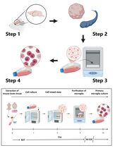
Primary microglia, in either mono-culture or co-culture with neurons or astrocytes, are a powerful tool for studying mechanisms underlying microglial inflammatory responses and cell type-specific interactions in the central nervous system (CNS). This protocol provides the details of how to prepare high purity primary microglia from newborn mouse pups. The overall steps include brain cell dissociation, mixed glial cell culture, and microglia isolation.
Background
In recent years, neuroinflammation has become a hotspot area in neuroscience studies. Inflammatory responses, such as glial activation and cytokine upregulation, were observed in brains of patients with various neurological diseases (Fan et al., 2015; Koshimori et al., 2015; Garden and Campbell, 2016). Neuroinflammation is considered not only a consequence of pathological changes in the brain but also a contributor to disease progression (Schwartz et al., 2013). In addition, the physiological functions of inflammatory pathways, the importance of which were previously underestimated, are being revealed as surprisingly versatile. For instance, activation of the complement signaling pathway is commonly observed in the central nervous system (CNS) in neurological diseases and is suspected to be involved in disease pathophysiology (Michailidou et al., 2015; Loeffler et al., 2008). Now we know that it also plays essential function in the developmental regulation of synaptic refinement (Stevens et al., 2007). Along with the increasing attention on inflammation, interest in microglial function during development, neuroprotection, and pathogenesis continues growing. Microglia are resident innate immune cells of myeloid lineage located in the brain and are critical components of the immune system in the CNS. The activation of microglia in some neurological diseases may directly participate in pathogenic processes. For instance, TREM2 mutations, which affects only microglia, are a genetic risk factor for Alzheimer’s disease (Yuan et al., 2016; Wang et al., 2015). At the same time, developmental roles of microglia are being revealed. For example, synaptic maturation during early development requires microglia and this regulation may underline the pathogenesis of developmental diseases such as autism (Edmonson et al., 2016; Stephan et al., 2012). Tools for studying microglia include in vivo models (e.g., microglia-deficient PU.1 knockout mice [McKercher et al., 1996]) and in vitro systems such as immortalized microglial cell lines and primary microglial culture. While in vivo tools are powerful for demonstrating systematic microglial function, in vitro tools are ideal for mechanistic characterization due to the easy manipulation of experimental factors. Compared to immortalized microglial cell lines, primary microglia better mimic in vivo microglial properties (Stansley et al., 2012). The altered gene expression upon stimulation may be better presented in primary microglia than in microglial cell lines (Stansley et al., 2012; Henn et al., 2009). Here we described a protocol for establishing high purity primary microglial culture derived from neonatal mice and the method has yielded robust results in our work (Lian et al., 2016). Dissociated cells are collected through enzymatic digestion of collected brains and seeded to grow mixed glial culture. Microglia growing on top of a confluent astrocyte layer are purified through mechanical tapping of mixed glial culture.
Materials and Reagents
- 15 ml centrifuge tubes (Corning, catalog number: 430052 )
- 50 ml centrifuge tubes (Corning, catalog number: 430290 )
- 12-well plates (Corning, Costar®, catalog number: 3737 )
- New born pups (mouse, P0-P2)
- Poly-D-lysine (PDL) (Sigma-Aldrich, catalog number: P6407-5MG )
- Ethanol
- Dulbecco’s modified Eagle medium (DMEM) (Thermo Fisher Scientific, GibcoTM, catalog number: 11995065 )
- Fetal bovine serum (FBS) (GE Healthcare, HycloneTM, catalog number: SH30088.03 )
- 10,000 U/ml penicillin-streptomycin (Pen/Strep) (Thermo Fisher Scientific, GibcoTM, catalog number: 15140122 )
- Hanks’ balanced salt solution (HBSS) (Thermo Fisher Scientific, GibcoTM, catalog number: 24020117 )
- 1 M HEPES buffer solution (Thermo Fisher Scientific, GibcoTM, catalog number: 15630080 )
- Glucose (Thermo Fisher Scientific, Fisher Scientific, catalog number: D16-3 )
- Trypsin, powder (Thermo Fisher Scientific, GibcoTM, catalog number: 27250018 )
- Trypsin inhibitor (Sigma-Aldrich, catalog number: T6522-100MG )
- Deoxyribonuclease I (DNase I) (Sigma-Aldrich, catalog number: DN25-100MG )
- Culture medium (500 ml) (see Recipes)
- Dissection medium (500 ml) (see Recipes)
- 2.5% trypsin (20 ml) (see Recipes)
- 1 mg/ml trypsin inhibitor (20 ml) (see Recipes)
- 10 mg/ml DNase (20 ml) (see Recipes)
Equipment
- Vented cap T-75 culture flask (Corning, catalog number: 3276 )
- Dissection tools
- Fine scissors (Fine Science Tools, catalog number: 14060-09 )
- Spring scissors (Fine Science Tools, catalog number: 15009-08 )
- Curved standard forceps (Fine Science Tools, catalog number: 11052-10 )
- Fine forceps (Fine Science Tools, catalog number: 11370-40 )
- Centrifuge machine (Eppendorf, model: 5702 )
- Hemocytometer
- Ventilation hood (Thermo Fisher Scientific, Thermo ScientificTM, catalog number: 1323 )
- CO2 cell culture incubator (Thermo Fisher Scientific, Thermo ScientificTM, catalog number: 50144906 )
- 37 °C water bath (Thermo Fisher Scientific, Thermo ScientificTM, model: TSGP02 )
Procedure
文章信息
版权信息
© 2016 The Authors; exclusive licensee Bio-protocol LLC.
如何引用
Readers should cite both the Bio-protocol article and the original research article where this protocol was used:
- Lian, H., Roy, E. and Zheng, H. (2016). Protocol for Primary Microglial Culture Preparation. Bio-protocol 6(21): e1989. DOI: 10.21769/BioProtoc.1989.
-
Lian, H., Litvinchuk, A., Chiang, A. C., Aithmitti, N., Jankowsky, J. L. and Zheng, H. (2016). Astrocyte-microglia cross talk through complement activation modulates amyloid pathology in mouse models of Alzheimer’s disease. J Neurosci 36(2): 577-589.
分类
神经科学 > 细胞机理 > 细胞分离和培养
细胞生物学 > 细胞分离和培养 > 细胞分离
您对这篇实验方法有问题吗?
在此处发布您的问题,我们将邀请本文作者来回答。同时,我们会将您的问题发布到Bio-protocol Exchange,以便寻求社区成员的帮助。