

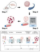
Microglial Phagocytosis Assay
小胶质细胞吞噬作用试验
发布: 2016年11月05日第6卷第21期 DOI: 10.21769/BioProtoc.1988 浏览次数: 19173
评审: Soyun KimPengpeng LiAnonymous reviewer(s)
参见作者原研究论文
The authors used this protocol in:
Jan 2016


Abstract
Phagocytosis is essential for microglial clearance of apoptotic cells, extracellular protein aggregates, and infectious bacteria in the central nervous system (CNS). While the preparation of primary microglial culture has been described elsewhere, this protocol describes the microglial phagocytosis experimental procedure and the subsequent measurement of microglial phagocytic ability using fluorescent latex beads or fluorescent amyloid beta 42 (Aβ42) peptides.
Background
Microglia play multiple roles in the central nervous system (CNS). Upon stimulation, microglia present complicated inflammatory responses including altered gene expression and morphological changes (Heneka et al., 2014; Cunningham, 2013). Cytokines are a critical cluster of proteins among the list of altered expression molecules by activated microglia. Through the potent signaling-capable cytokine receptors expressed on astrocytes, neurons, and other brain cell types, microglia communicate, recruit, and coordinate inflammatory events (Smith et al., 2012). Besides cytokine secretion, phagocytosis, which involves morphological changes in microglia, also adds to their role as guardians of environmental homeostasis within the CNS milieu. Microglial phagocytosis of pathogens, extracellular protein aggregates, and apoptotic cell debris dampens inflammation and protects neurons (Fu et al., 2014). Apart from pathogenic conditions, microglial phagocytosis is also involved in CNS development and synaptogenesis through eliminating nonfunctional synapses. Deficient or excess microglial phagocytic ability could lead to abnormal synaptic connections and deposits of aggregated proteins (Schafer et al., 2012; Lian et al., 2016). Here we describe a protocol for measuring microglial phagocytic ability using in vitro cultured primary microglia. To mimic exogenous particles and protein aggregates, we used latex beads and amyloid β protein as the substrates for microglia to engulf.
Materials and Reagents
- 24-well plates (Corning, Costar®, catalog number: 3527 )
- 12 mm glass coverslips (Thermo Fisher Scientific, Fisher Scientific, catalog number: 12-545-81 )
- 1.5 ml centrifuge tubes (Corning, Axygen®, catalog number: MCT-150-R )
- Fluorescent latex beads of 1 μm diameter (Sigma-Aldrich, catalog number: L1030 )
- Poly-D-lysine (PDL) (Sigma-Aldrich, catalog number: P6407-5MG )
- Distilled water (Thermo Fisher Scientific, GibcoTM, catalog number: 15230147 )
- Dulbecco’s modified Eagle medium (DMEM) (Thermo Fisher Scientific, GibcoTM, catalog number: 11995065 )
- Fetal bovine serum (FBS) (GE Healthcare, HycloneTM, catalog number: SH30088.03 )
- FAM-labelled Aβ42 peptides (AnaSpec, catalog number: AS-23526-01 )
- Dimethyl sulfoxide (DMSO) (Sigma-Aldrich, catalog number: 472301 )
- Phosphate buffered salt (PBS)
- 4% PFA (Santa Cruz Biotechnology, catalog number: sc-281692 )
- Mounting medium with DAPI (Vector Laboratories, catalog number: H-1200 )
- Microglial culture media (500 ml) (see Recipes)
Equipment
- Ventilation hood (Thermo Fisher Scientific, Thermo ScientificTM, catalog number: 1323 )
- CO2 cell culture incubator (Thermo Fisher Scientific, Thermo ScientificTM, catalog number: 50144906 )
- 37 °C water bath (Thermo Fisher Scientific, Thermo ScientificTM, model: TSGP02 )
- Cell counter
Software
- ImageJ (http://fiji.sc/)
Procedure
文章信息
版权信息
© 2016 The Authors; exclusive licensee Bio-protocol LLC.
如何引用
Readers should cite both the Bio-protocol article and the original research article where this protocol was used:
- Lian, H., Roy, E. and Zheng, H. (2016). Microglial Phagocytosis Assay. Bio-protocol 6(21): e1988. DOI: 10.21769/BioProtoc.1988.
- Lian, H., Litvinchuk, A., Chiang, A. C., Aithmitti, N., Jankowsky, J. L. and Zheng, H. (2016). Astrocyte-microglia cross talk through complement activation modulates amyloid pathology in mouse models of Alzheimer's disease. J Neurosci 36(2): 577-589.
分类
神经科学 > 细胞机理 > 细胞分离和培养
细胞生物学 > 细胞分离和培养 > 细胞分离
您对这篇实验方法有问题吗?
在此处发布您的问题,我们将邀请本文作者来回答。同时,我们会将您的问题发布到Bio-protocol Exchange,以便寻求社区成员的帮助。