

Organotypic Spinal Cord Slice Cultures and a Method to Detect Cell Proliferation in These Slices
器官型脊髓片培养和脊髓片上细胞增殖的检测方法


发布: 2016年10月05日第6卷第19期 DOI: 10.21769/BioProtoc.1951 浏览次数: 12710
评审: Manuel D. GaheteKae-Jiun ChangAnonymous reviewer(s)
参见作者原研究论文
The authors used this protocol in:
Sep 2015


Abstract
In these culture models, the normal cytoarchitecture and local neuronal circuits of the spinal cord are preserved, offering a compromise between dissociated cell cultures and complete animal studies. The addition of 5-ethynyl-2’-deoxyuridine (EdU) to the culture medium allows for the detection of proliferating cells.
Keywords: Neurogenesis (神经发生)Materials and Reagents
- Filter paper, 55 mm (Sigma-Aldrich, Whatman®, catalog number: WHA1441055 )
- Millicell organotypic filter inserts 0.4 µm, 30 mm (EDM Millipore, catalog number: Picmorg50 )
- 6-well culture dishes (Thermo Fisher Scientific, Thermo ScientificTM, catalog number: 140675 )
- 24-well histology trays (Sigma-Aldrich, CELLSTAR®, catalog number: 662160 )
- 50 mm sterilin Petri dish (Camlab, catalog number: p17118 )
Note: This link leads to a range of Petri dishes. - Pipette tips, 1,000 µl (SARSTEDT, catalog number: 70.762.010 )
- Pipette tips, 200 µl (SARSTEDT, catalog number: 70.760.012 )
- Syringe
- Glass microscope slides (Academy Science, catalog number: N/A143 )
- Glass coverslips (VWR, catalog number: 631-0133 )
- Tin foil
- Mice (8-20 days old)
- Ethanol (Sigma-Aldrich, catalog number: 32221 )
- Acetone (Alfa Aesar, catalog number: L10407 )
- Sodium pentobarbitone (Pentoject) (Animalcare, catalog number: XVD-132 )
- 5-ethynyl-2’-deoxyuridine (EdU) (Thermo Fisher Scientific, Molecular ProbesTM, catalog number: A10044 )
- Paraformaldehyde (Sigma-Aldrich, catalog number: 158127 )
- Phosphate buffered saline (Dulbecco A) (PBS) (Oxoid Limited, catalog number: BR0014 )
- Triton X-100 (VWR, catalog number: 28817.295 )
- Copper sulphate pentahydrate (CuSO4·5H2O) (VWR, catalog number: 84845.230 )
- Biotin-azide (Kerafast, catalog number: EVU101 )
- Ascorbic acid (Thermo Fisher Scientific, Fisher Scientific, catalog number: 12950364 )
- Streptavidin Alexa555 conjugate (Thermo Fisher Scientific, Molecular ProbesTM, catalog number: S-32355 )
- Vectashield mounting medium (Vecta Laboratories, catalog number: H-1000 and H-1200 )
- DAPI (4’,6-diamidino-2-phenylindole)
- Nail lacquer
- Anti-PKD2L-1 (polycystic kidney disease 2-like 1 protein)
- PBS tablets
- Sodium phosphate monobasic monohydrate (NaH2PO4·H2O) (Thermo Fisher Scientific, Fisher Scientific, catalog number: 10667823 )
- Sodium phosphate dibasic anhydrous (Na2HPO4) (Thermo Fisher Scientific, Fisher Scientific, catalog number: 10396313 )
- Trizma base (Sigma-Aldrich, catalog number: T1503 )
- Trizma hydrochloride (Sigma-Aldrich, catalog number: T3253 )
- Sucrose (Thermo Fisher Scientific, Fisher Scientific, catalog number: 10386100 )
- Sodium bicarbonate (NaHCO3) (Sigma-Aldrich, catalog number: 31437 )
- Potassium chloride (KCl) (Sigma-Aldrich, catalog number: 746436 )
- Magnesium sulphate (MgSO4·7H2O) (VWR, catalog number: 25165.260 )
- Glucose (Thermo Fisher Scientific, Fisher Scientific, catalog number: 10539380 )
- Calcium chloride (CaCl2) (Sigma-Aldrich, catalog number: 21114 )
- Dulbecco’s modified Eagle’s medium (DMEM) (Sigma-Aldrich, catalog number: D6546 )
- Penicillin and streptomycin (Sigma-Aldrich, catalog number: P4458 )
- Neurobasal A medium (Thermo Fisher Scientific, GibcoTM, catalog number: 10888-022 )
- L-glutamine (Sigma-Aldrich, catalog number: G7513 )
- B-27 supplement (Thermo Fisher Scientific, GibcoTM, catalog number: 17504-044 )
- Fetal bovine serum (FBS) (Sigma-Aldrich, catalog number: F6178 )
- Phosphate buffered saline (PBS) (see Recipes)
- 0.2 M phosphate buffer (PB) (see Recipes)
- 0.1 M Tris buffer (see Recipes)
- Sucrose artificial cerebrospinal fluid (see Recipes)
- 8% paraformaldehyde solution (PFA) (see Recipes)
- 4% paraformaldehyde solution in 0.1 M PB (see Recipes)
- Dissection medium (see Recipes)
- Culture medium with serum (see Recipes)
- Culture medium serum free (see Recipes)
Equipment
- Autoclave (Dixons Surgical Instrument, model: VARIO 1528 )
- P1000 pipetman classic pipette (Gilson Scientific, catalog number: F123602 )
- P20 pipetman classic pipette (Gilson Scientific, catalog number: F123600 )
- Laminar flow hood (biological safety cabinet) (The Baker Company, model: Steril Gard class II type A )
- CO2 incubator (Panasonic, model: MCO-18AC-PE )
- Dissection microscope (Vickers, instruments)
- Dissection equipment (Figure 1)
- Dissection scissors
- Spring scissors
- Fine forceps x 2
- Microspatula
- Tissue chopper (McIlwain)
- Razor blades (Wilkinson sword)
- Shaker plate (IKA, model: Vibrax-vxr )
- Fine paint brush
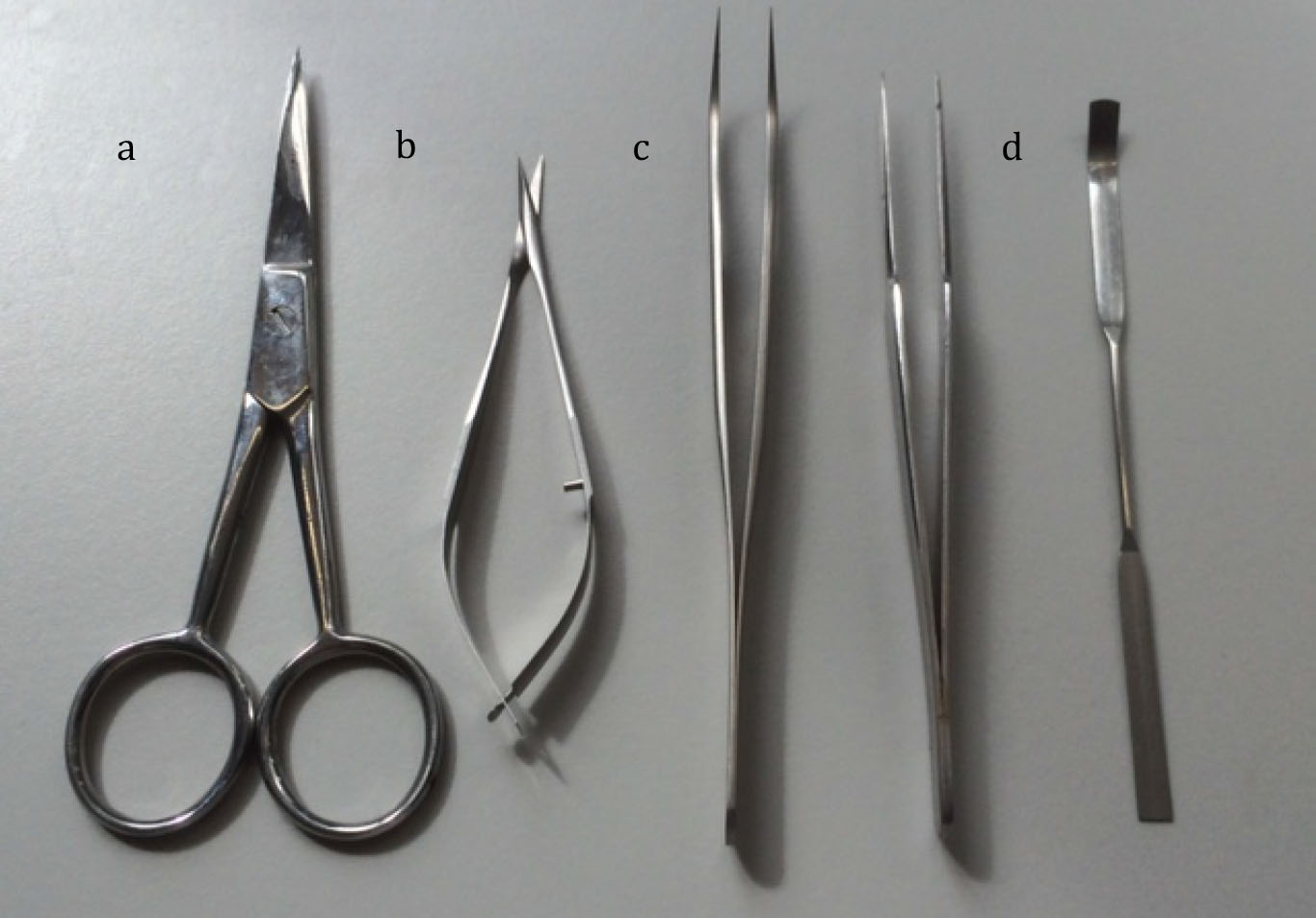
Figure 1. Equipment used for dissection
Procedure
文章信息
版权信息
© 2016 The Authors; exclusive licensee Bio-protocol LLC.
如何引用
Daniel, J. M., Deuchars, J. and Deuchars, S. A. (2016). Organotypic Spinal Cord Slice Cultures and a Method to Detect Cell Proliferation in These Slices . Bio-protocol 6(19): e1951. DOI: 10.21769/BioProtoc.1951.
分类
神经科学 > 细胞机理 > 细胞分离和培养
细胞生物学 > 细胞分离和培养 > 细胞生长
您对这篇实验方法有问题吗?
在此处发布您的问题,我们将邀请本文作者来回答。同时,我们会将您的问题发布到Bio-protocol Exchange,以便寻求社区成员的帮助。