

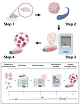
Preparation of Single Cell Suspensions from Mouse Aorta
从小鼠主动脉制备单细胞悬浮液


发布: 2016年06月05日第6卷第11期 DOI: 10.21769/BioProtoc.1832 浏览次数: 18383
评审: Ruth A. FranklinAnonymous reviewer(s)
参见作者原研究论文
The authors used this protocol in:

Jun 2015
Advertisement


Abstract
Atherosclerosis is a chronic inflammatory disease of the arterial wall characterized by lipid deposition, plaque formation, and immune cell infiltration. Innate and adaptive immune cells infiltrate the artery during development of the disease. Moreover, advanced disease leads to formation of artery tertiary lymphoid organs in the adventitia (Grabner et al., 2009; Hu et al., 2015). Various and diverse types of immune cells have been identified in the aorta adventitia vs atherosclerotic plaques (Elewa et al., 2016; Galkina et al., 2006; Lotzer et al., 2010; Mohanta et al., 2016; Mohanta et al., 2014; Moos et al., 2005; Srikakulapu et al., 2016; Zhao et al., 2004). There are conflicting reports on the number and subtypes of immune cells in the aorta depending on the age of the animals, the protocol that is used to obtain single cell suspensions, and the dietary conditions of the mice (Campbell et al., 2012; Clement et al., 2015; Galkina et al., 2006; Kyaw et al., 2012). The number of immune cells in the aorta differs as much as tenfold using different protocols (Butcher et al., 2012; Galkina et al., 2006; Gjurich et al., 2015; Grabner et al., 2009; Hu et al., 2015). These discrepant results call for a protocol that robustly documents bona fide aorta cells rather than those in the surrounding tissues or blood. Critical methodological hurdles include the removal of adjacent adipose tissue and small paraaortic lymph nodes lining the entire aortic tree that are not visible by the naked eye. A dissection microscope is therefore recommended. Moreover protocols of aorta preparations should ascertain that lymphocyte aggregates referred to as fat associated lymphoid clusters (FALCs) (Benezech et al., 2015; Elewa et al., 2015) that are often present at the border between the adipose tissue and the adventitia are removed before enzyme digestion. We propose - besides other approaches (Hu et al., 2015; Mohanta et al., 2014) - a combination of immunohistochemical staining and fluorescence activated cell sorter (FACS) analyses from single cell suspensions to quantify the cells of interest. This protocol describes isolation of single cells from mouse aorta for FACS and other analysis.
Materials and Reagents
- 50 ml Falcon tube (VWR International, CellStar®, catalog number: 188271 )
- 100 µm cell strainer (BD, catalog number: 352360 )
Note: Currently, it is “Corning, Falcon®, catalog number: 352360”. - 1 ml syringe (Henke-Sass, Wolf GmbH, Soft-JECT®, catalog number: 5010-200V0 )
- 5 ml syringe (BD, catalog number: 309646 )
- Needle-26G (B. Braun Medical Inc., catalog number: 4657683 )
- 6-well plate (BD Falcon, catalog number: 353046 )
Note: Currently, it is “Corning, Falcon®, catalog number: 353046”. - 1.5 ml Eppendorf tube (Eppendorf AG, catalog number: 0030123328 )
- Trypan blue solution (Sigma-Aldrich, catalog number: 93595 )
- Phosphate-buffered saline (PBS), pH 7.4 (Thermo Fisher Scientific, GibcoTM, catalog number: 10010023 )
- Dulbecco’s phosphate-buffered saline (DPBS) (Thermo Fisher Scientific, GibcoTM, catalog number: 14040133 )
- Fetal bovine serum (FBS) (PAN Biotech UK Ltd., catalog number: P30-1506 )
- Ethylenediaminetetraacetic acid (EDTA) (Sigma-Aldrich, catalog number: E6758 )
- Collagenase from Clostridium histolyticum, type I (Sigma-Aldrich, catalog number: C0130 )
- Collagenase from Clostridium histolyticum, type XI (Sigma-Aldrich, catalog number: C7657 )
- Hyaluronidase from bovine testes, type I-s (Sigma-Aldrich, catalog number: H3506 )
- DNase I (Sigma-Aldrich, catalog number: 11284932001 )
- 4-(2-Hydroxyethyl)-1-piperazineethanesulfonic acid (HEPES) (1 M) (Thermo Fisher Scientific, GibcoTM, catalog number: 15630106 )
- Ethanol solution (Sigma-Aldrich, catalog number: 48075 )
- Anti-mouse CD45 APC antibody (Thermo Fisher Scientific, eBioscience, catalog number: 17-0451-82 )
- LIVE/DEAD® fixable blue dead cell stain kit (Invitrogen, catalog number: L23105 )
Note: Currently, it is “Thermo Fisher Scientific, Molecular ProbesTM, catalog number: L23105”. - Fc block (anti-CD16/32) (Thermo Fisher Scientific, eBioscience, catalog number: 16-0161-82 )
- FACS buffer (see Recipes)
- EDTA buffer (see Recipes)
- Enzyme cocktail (see Recipes)
Equipment
- Dissecting scissors (Fine Science Tools, catalog number: 91460-11 )
- Curved forceps (Fine Science Tools, catalog number: 11073-10 )
- CO2 supply machine (Next Advance, model: Quietek CO2 induction system )
- Neubauer cell counting chamber (Marienfeld-Superior)
- Microscope (Carl Zeiss Microscopy, model: Axiovert 40C )
- Dissecting microscope equipped with cold light (Carl Zeiss Microscopy, model: Stemi2000 )
- Water bath (Thomas Scientific, model: 1196x11 )
- BD LSRFortessa (BD Bioscience)
Procedure
文章信息
版权信息
© 2016 The Authors; exclusive licensee Bio-protocol LLC.
如何引用
Hu, D., Yin, C., Mohanta, S., Weber, C. and Habenicht, A. J. R. (2016). Preparation of Single Cell Suspensions from Mouse Aorta. Bio-protocol 6(11): e1832. DOI: 10.21769/BioProtoc.1832.
分类
免疫学 > 免疫细胞分离 > 白细胞
细胞生物学 > 细胞分离和培养 > 细胞分离
您对这篇实验方法有问题吗?
在此处发布您的问题,我们将邀请本文作者来回答。同时,我们会将您的问题发布到Bio-protocol Exchange,以便寻求社区成员的帮助。