

Proximity Ligation Assay (PLA) Protocol Using Duolink® for T Cells
用基于PLA原理的 Duolink®体系观察T淋巴细胞中蛋白相互作用的方法


发布: 2016年05月20日第6卷第10期 DOI: 10.21769/BioProtoc.1811 浏览次数: 20588
评审: Jia LiGriselda Zuccarino-CataniaAnonymous reviewer(s)
参见作者原研究论文
The authors used this protocol in:
Jul 2015
Advertisement


Abstract
Protein-protein interaction experiments, such as co-immunoprecipitation (IP) assays, classically require tremendous amount of cells. This becomes a problem when your work focuses on rare cell populations (e.g., lymphocyte subtypes). O-link Bioscience has developed Proximity Ligation Assay (PLA) reagents and procedures to alleviate and solve this kind of issue. Moreover PLA experiments are read out using fluorescence or bright field microscopy, providing additional information on intracellular interactions localization significantly bettering classical IP procedures.
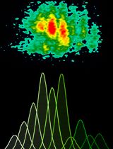
PLA reagents are made of complementary small oligonucleotides “minus” and “plus” probes which specifically recognize host species from the primary antibodies (Abs) targeting the two proteins you are interested in. Experiments have to be designed with primary Abs from different species (rabbit, mouse or goat) as PLA probes “minus” or “plus” react against a specific host species of the primary antibody (e.g. “plus” anti-rabbit with “minus” anti-mouse or “plus” anti-mouse with “minus” anti-rabbit combos are allowed if mouse and rabbit primary Abs are used). When the two PLA probes are close enough (40 nm) a ligation occurs upon ligase incubation generating a circle DNA. These circle-forming DNAs are next amplified thanks to a polymerase and complementary fluorescent nucleotides, being incorporated at this step. Each luminescent spot is thereafter considered being an interaction site between the two proteins (Figure 1).
Figure 1. Schematic representation of Duolink® experiment. Primary Abs from different host species were used [i.e., Mouse (Ms) anti-Nlrp3 and Rabbit (Rb) anti-IRF4]. When protein-protein interaction occurs PLA probes allow incorporation of fluorescent oligonucleotides which are analyzed by microscopy.
PLA experiments have been performed on differentiated T cells from mice. T cells were grown on cover slip coated with poly-L-Lysine. Please be aware that for T cells and other non-adherent cells, experiments and staining are also possible in 500 µl microtubes until the last washing step before mounting cover slip on slide. This microtube method saves cytokines reagents and allows T cells to grow with usual methods but centrifugation repetition for washing steps is hazardous. Primary Abs incubation also requires also a specific setup if microtube method is chosen.
Materials and Reagents
- Glass cover slip (Ø13 mm thickness, No. 1, 5) (VWR International, catalog number: 631-0150 )
- Primary antibodies (Abs)
- Mouse anti-Nlrp3 (1/100) (Adipogen International, catalog number: AG-20B-0014 )
Note: Starting concentration of mouse anti-Nlrp3 is1 µg/µl. - Rabbit anri-IRF4 (1/100) (Novus Biologicals, catalog number: NBP1-00893 )
Note: Starting concentration of rabbit anri-IRF4 is 0.08 µg/µl.
- Mouse anti-Nlrp3 (1/100) (Adipogen International, catalog number: AG-20B-0014 )
- Duolink® reagents
- Anti-rabbit PLUS probe (1/5) (Sigma-Aldrich, catalog number: Duo92002 )
- Anti-mouse MINUS probe (1/5) (Sigma-Aldrich, catalog number: Duo92004 )
- Ligation reagents (5x) (Sigma-Aldrich, catalog number: Duo92007 )
Note: Ligation reagents, ligase and amplification reagents are all included in the pack # DUO92007 . - Ligase (1 unit/μl) (Sigma-Aldrich, catalog number: Duo82029 )
- Amplification reagents (containing orange labeled oligonucleotides) (5x) (Sigma-Aldrich, catalog number: Duo92007 )
- Polymerase (10 unit/μl) (Sigma-Aldrich, catalog number: Duo82030 )
- Wash buffer A (Sigma-Aldrich, catalog number: Duo82049 )
- Wash buffer B (Sigma-Aldrich, catalog number: Duo82049 )
- Anti-rabbit PLUS probe (1/5) (Sigma-Aldrich, catalog number: Duo92002 )
- Poly-L-Lysine (Sigma-Aldrich, catalog number: P4707 )
- Triton X-100 (0.1% in PBS) (Sigma-Aldrich, catalog number: T8787 )
- Blocking & Abs dilution buffer (0.5% BSA in PBS)
- Mounting medium containing DAPI (Thermo Fisher Scientific, Molecular ProbesTM, catalog number: P36931 )
- 1x PBS (Lonza, catalog number: 17-516F )
- RNase free water (Thermo Fisher Scientific, InvitrogenTM, catalog number: 10977035 )
- 4% formaldehyde (PFA) (VWR International, catalog number: 9713.1000 )
Equipment
- Pipets (from 1 to 1,000 µl)
- Fluorescence microscope (with DAPI and Cy2 or Cy3 emission filter) (ZEISS, model: Imager M2)
Note: Objective 63x is recommended for T cells imaging. - 37 °C incubator
- Freeze block for enzymes
- Orbital shaker
Software
- ImageJ
Procedure
文章信息
版权信息
© 2016 The Authors; exclusive licensee Bio-protocol LLC.
如何引用
Derangère, V., Bruchard, M., Végran, F. and Ghiringhelli, F. (2016). Proximity Ligation Assay (PLA) Protocol Using Duolink® for T Cells. Bio-protocol 6(10): e1811. DOI: 10.21769/BioProtoc.1811.
分类
免疫学 > 免疫细胞功能 > 综合
您对这篇实验方法有问题吗?
在此处发布您的问题,我们将邀请本文作者来回答。同时,我们会将您的问题发布到Bio-protocol Exchange,以便寻求社区成员的帮助。