

Preparation and Immunofluorescence Staining of the Trachea in Drosophila Larvae and Pupae
果蝇幼虫和蛹中气管的制备和免疫荧光染色
发布: 2016年05月05日第6卷第9期 DOI: 10.21769/BioProtoc.1797 浏览次数: 21769
评审: Xuecai GeJihyun KimAnonymous reviewer(s)
参见作者原研究论文
The authors used this protocol in:
Jan 2014
Advertisement


Abstract
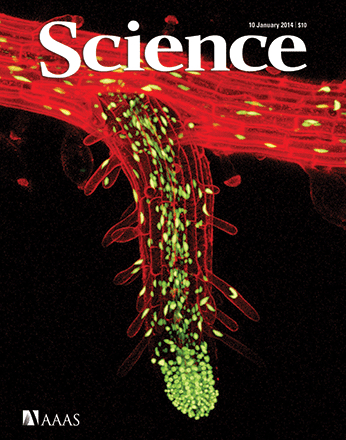
The Drosophila melanogaster trachea is a branched network of rigid chitin-lined tubes that ramify throughout the body and functions as the fly’s respiratory organ. Small openings at the ends of the tracheal tubes allow gas exchange to occur by diffusion between internal tissues and the exterior environment. Tracheal tubes are lined by a single layer of epithelial cells, which secrete chitin and control tube morphology and size. Studies of tracheal development in Drosophila embryos have elucidated fundamental mechanisms of tube morphogenesis and maintenance in vivo, and identified major signaling pathways that regulate these processes (Manning and Krasnow, 1993; Affolter and Shilo, 2000; Zuo et al., 2013; Kerman et al., 2006; Schottenfeld et al., 2010). In recent years, there has been growing interest in the trachea during metamorphosis, when tracheal branches that had served as the respiratory organ in the larva decays and is repaired or replaced by new tracheal tissue arising from committed tracheal progenitor cells, or mature tracheal cells de-differentiated to a progenitor state (Manning and Krasnow, 1993; Sato and Kornberg, 2002; Guha et al., 2008; Guha, and Kornberg, 2005; Weaver and Krasnow, 2008; Pitsouli and Perrimon, 2010; Chen and Krasnow, 2014) forming the adult tracheal by the end of the process. The ongoing decay and tissue formation models aspects of tissue repair and regeneration in other organisms, and has been used to understand how progenitor cells divide and differentiate (Pitsouli and Perrimon, 2010; Pitsouli and Perrimon, 2013), and how they grow out of their niche to replace decaying tissue (Chen and Krasnow, 2014). Here, we present a protocol to dissect, fix, and immunostain tracheal tissue in Drosophila larvae and pupae undergoing metamorphosis. This protocol can be used to immunostain proteins expressed in tracheal tissue, or to amplify signals from weakly expressed fluorescent reporters (as shown in Figure 6). With the appropriate antibodies and genetic reporters, this protocol can be used to visualize decaying larval trachea and the progenitor cells that replace them in a time-course analysis, as well as determine expression of proteins in these cells that may play a role in tissue decay and replacement.
Materials and Reagents
- 60 mm x 15 mm petri dish [i.e., Falcon® Petri dish (Corning, catalog number: 351007 )]
- Flat-bottom 4-well dish [i.e., Nunclon® Δ Multidishes, 4 wells, flat bottom (Sigma-Aldrich, catalog number: D6789-1CS )]
- Gold SealTM Rite-OnTM Frosted microslides (VWR International, Erie Scientific, catalog number: 3050 )
- Micro cover glasses (coverslips), 22 x 22 mm Square No. 1 (VWR International, catalog number: 48366-067 )
- Black electrical tape (i.e., 3M Scotch Super 33+ Vinyl Electrical Tape 0.75 in x 450 in)
- Clear nail polish [i.e., crystal clear (Sally Hansen Hard as nails polish)]
- KimwipesTM (4.4 x 8.4 in.) (Thermo Fisher Scientific, catalog number: 06-666 )
- Austerlitz Insect Pins® ,12 mm length x 0.10 mm diameter (Minutiens in stainless steel, size 0.10 mm) (Entomoravia)
- 10 μl, 200 μl and 1,000 μl pipet tips (USA Scientific, TipOne, catalog number: 1111-3000 , 1111-0000 and 1111-2021 )
- Disposable glass Pasteur pipettes (Corning, catalog number: 7095D-5x ) with 1 ml rubber bulbs (Sigma-Aldrich, catalog number: Z111589 )
- Aluminum foil
- Parafilm M® All-Purpose laboratory film (2" x 250') (VWR International, Bemis Company, catalog number: PM992 )
- Drosophila melanogaster larvae and pupae of desired genotype raised at 25 °C
- Vials and bottles with closures [FisherbrandTM stock bottles (catalog number: AS117 ), FisherbrandTM cotton balls (catalog number: 22-456-880 ), FisherbrandTM Drosophila products, BuzzPlugsTM (catalog number: AS277 ), FisherbrandTM Drosophila vials (catalog number: AS514 )] with Drosophila food (see Cold Spring Harbor Protocols, 2014)
- Dow Corning SYLGARD® 184 Silicone Elastomer Kit [184 SIL ELAST KIT 0.5 KG (Ellsworth Adhesives)]
- 4% paraformaldehyde (PFA) diluted in PBS [i.e., 16% paraformaldehyde (VWR International, catalog number: 100503-916 ) diluted to 4% in PBS]
- Primary antibody to stain protein of interest [i.e., Chicken-anti-GFP (Abcam, catalog number: ab13970 ) to stain tracheal-expressed GFP in ppk4-Gal4, UAS-GFP larvae and pupae]
- Fluorescence conjugated secondary antibody to visualize and amplify primary antibody staining [i.e., Alexa488-conjugated Goat-anti-Chicken (Thermo Fisher Scientific, InvitrogenTM, catalog number: A-11039 ) to stain the above anti-GFP primary antibody]
- Normal serum from the same species as the secondary antibody [i.e., normal goat serum (Vector laboratories, catalog number: S-1000 )]
- Vectashield® mounting media (Vector Laboratories, catalog number: H-1000 )
- NaCl
- KCl
- Na2HPO4
- KH2PO4
- 1x Phosphate-buffered saline (PBS) (see Recipes)
- TritonTM X-100 (Sigma-Aldrich, catalog number: X100 ) diluted to 0.1% in PBS (see Recipes)
- Block solution (see Recipes)
- DAPI staining solution (see Recipes)
- Dissection dish (see Recipes)
Equipment
- Incubator to house Drosophila set to 25 °C
- Stereomicroscope with light source [i.e., Carl ZeissTM StemiTM 2000C with KL 300 LED Cold Light Source (120 V) (Thermo Fisher Scientific, catalog number: 12-070-284 )]
- Dumont #5 mirror finish forceps biology tips/straight/inox/11 cm (Fine Science Tools, catalog number: 11252-23 )
- Vannas spring scissors-straight/sharp/8 cm/3 mm cutting edge (Fine Science Tools, catalog number: 15000-00 )
- Benchtop shaker (i.e., Bellco Glass 7744-06115 Mini Orbital Shaker)
- Clay AdamsTM Nutator Mixer (BD, catalog number: 421105 )
- P2, P20, P100, and P1000 Pipetman® Pipettes (Gilson Scientific Ltd., catalog number: F144801 , F123600 , F123615 and F123602 )
- Small watercolor paintbrush, round, size 0
- Stainless steel spatula with a micro spoon end (Ted Pella Inc., catalog number: 13500 )
Procedure
文章信息
版权信息
© 2016 The Authors; exclusive licensee Bio-protocol LLC.
如何引用
Chen, F. (2016). Preparation and Immunofluorescence Staining of the Trachea in Drosophila Larvae and Pupae. Bio-protocol 6(9): e1797. DOI: 10.21769/BioProtoc.1797.
分类
发育生物学 > 形态建成 > 变态发育
细胞生物学 > 组织分析 > 组织染色
您对这篇实验方法有问题吗?
在此处发布您的问题,我们将邀请本文作者来回答。同时,我们会将您的问题发布到Bio-protocol Exchange,以便寻求社区成员的帮助。