

Isolation and Culture of Human CD133+ Non-adherent Endothelial Forming Cells
分离和培养人 CD133+非粘连内皮形成细胞
发布: 2016年04月05日第6卷第7期 DOI: 10.21769/BioProtoc.1772 浏览次数: 11529
评审: Ningfei AnThomas J. BartoshAnonymous reviewer(s)
参见作者原研究论文
The authors used this protocol in:
May 2015
Advertisement


Abstract
Circulating endothelial progenitor cells (EPCs) have been the focus of many clinical trials due to their roles in revascularisation following ischemic events such as acute myocardial infarction as well as their contribution to vascular repair during organ transplantation. Research on EPCs has been controversial due to the lack of distinct markers expressed at the cell surface and varying methods for isolation and culture have resulted in the identification of a multitude of cell types, with differing phenotype and function, all falling under the label of “EPCs”. The most widely documented EPCs isolated for cell therapy are adherent in nature and lacking the progenitor markers such as CD133 and therefore unlikely to represent a true circulating EPC, the cells mobilised in response to a vascular injury.
We recently published the isolation and extensive characterisation of a population of non-adherent endothelial forming cells (naEFCs) (Appleby et al., 2012) (Figure 1). These cells expressed the progenitor cell markers (CD133, CD34, CD117, CD90 and CD38) together with mature endothelial cell markers (VEGFR2, CD144 and CD31). These cells also expressed low levels of CD45 but did not express the lymphoid markers (CD3, CD4, CD8) or myeloid markers (CD11b and CD14) which distinguishes them from ‘early’ EPCs, the ‘late outgrowth EPC’ [more recently known as endothelial colony forming cells (ECFCs)] as well as mature endothelial cells (ECs). Figure 2A exemplifies the surface expression profile of the naEFCs. Functional studies demonstrated that these naEFCs (i) bound Ulex europaeus lectin (Figure 2A), (ii) demonstrated acetylated-low density lipoprotein uptake, (iii) increased vascular cell adhesion molecule (VCAM-1) surface expression in response to tumor necrosis factor and (iv) in co-culture with mature ECs increased the number of tubes, tubule branching and loops in a 3-dimensional in vitro matrix. More importantly, naEFCs placed in vivo generated new lumen containing vasculature lined by CD144 expressing human ECs and have contributed to various advances in scientific knowledge (Appleby et al., 2012; Barrett et al., 2011; Moldenhauer et al., 2015; Parham et al., 2015). Here, we describe the isolation and enrichment of a non-adherent CD133+ endothelial forming population of cells from human cord blood.
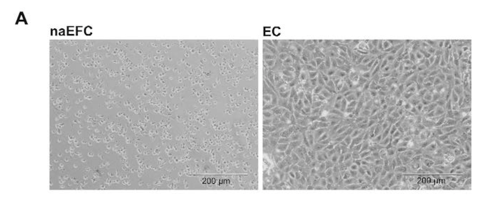
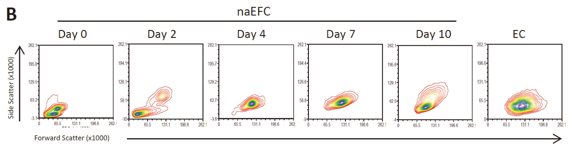
Figure 1. Enrichment of human naEFCs. A. Umbilical cord blood derived CD133+ enriched cells (naEFCs) at 4 days of culture and human umbilical vein endothelial cells (ECs) were compared for cell size by light microscopy. Scale bar=200 µm B. The cells were assessed for heterogeneity of enrichment process (0-10 days) via forward scatter and side scatter profiling using flow cytometric analysis and compared to mature ECs.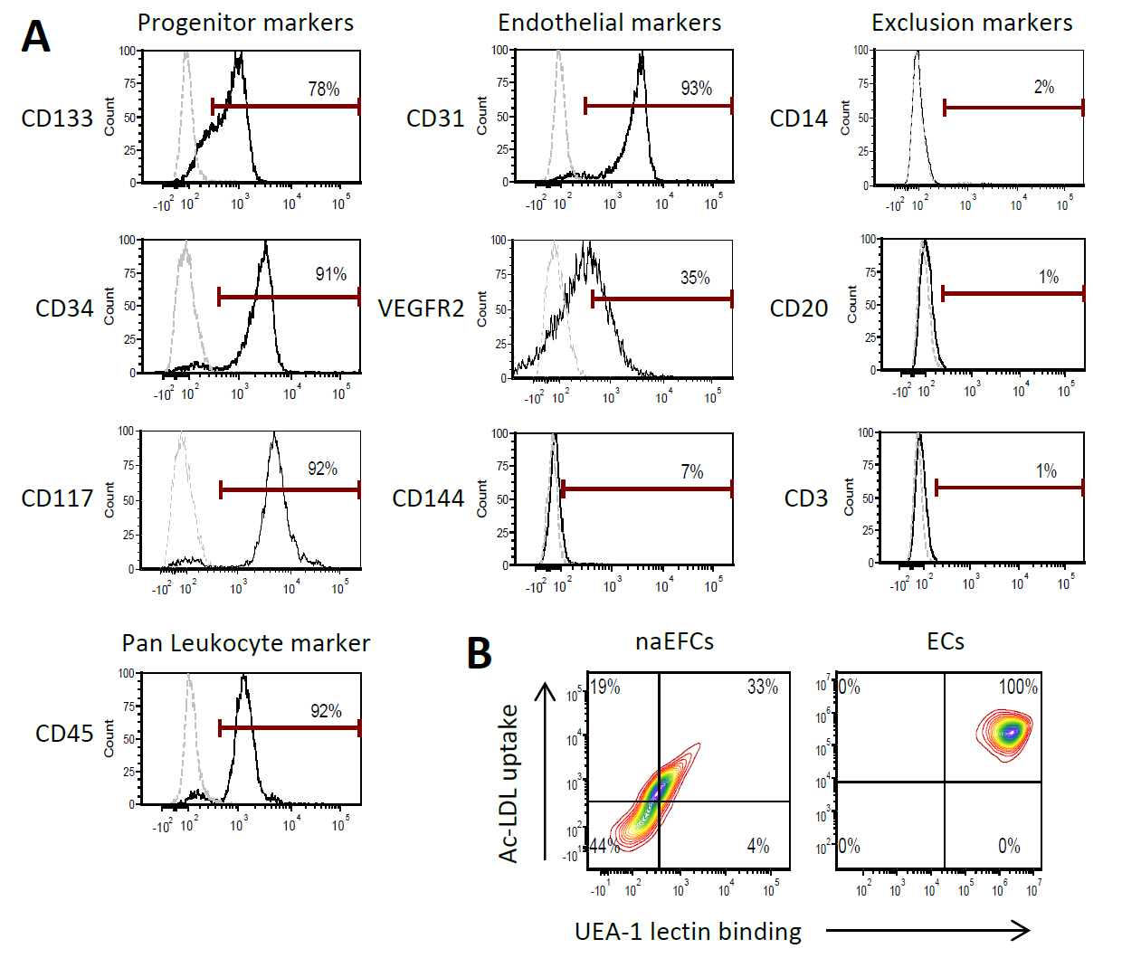
Figure 2. Surface expression phenotype of human naEFCs. A. CD133+ enriched cells at 4 days of culture were assessed for progenitor and endothelial markers by flow cytometry. Histograms show a representative experiment from ≥3 biological replicates where grey dashed lines represent isotype controls and solid black lines represent cells stained with the indicated marker. B. The function of the naEFCs was assessed by flow cytometry and compared to mature ECs, detecting the ability of cells to uptake DiI labelled acetylated low density lipoprotein (Ac-LDL) and bind FITC labelled Ulex europaeus agglutinin I (UEA-1) lectin. Density plots represent stained cells of one representative experiment from ≥3 biological replicates.
Materials and Reagents
- For manual cell sorting
MS Columns (Miltenyi Biotec, catalog number: 130-042-201 ) - MacoPharma cord blood collection bags (MacoPharma, catalog number: MSC1201DU )
- 50 ml tubes (Corning, Falcon®, catalog number: 352070 )
- 10 ml tubes (Sarstedt AG, catalog number: 62.9924.284 )
- 2 ml soft sterile bulb transfer pipette, sterile (Stephen Gould corporation, catalog number: 222-1S )
Note: Currently, it is “Capitol Scientific, catalog number: 222-1S”. - 20 µm filter (Sartorius Stedim Biotec, Minisart, catalog number: 16534-K )
- 24-well plate (Corning, Falcon®, catalog number: 353047 )
- Microfuge tubes
- 20 ml syringes
- 0.2 µm filter
- Human umbilical cord blood (40-250 ml)
- 20x Dulbecco’s phosphate buffered (DPBS) (Life Technologies, Gibco®, catalog number: 14200-075 )
Note: Currently, it is “Thermo Fisher Scientific, GibcoTM, catalog number: 14200-075”. - Sterile water (Baxter, catalog number: UKF7114 )
- LymphoprepTM (Axis-Shield, catalog number: 114547 )
- CD133 microbeads including human FcR blocking reagent (Miltenyi Biotec, catalog number: 130-050-801 )
- AutoMACS Pro Washing Solution (Miltenyi Biotec, catalog number: 130-092-987 )
- AutoMACS Running Buffer (Miltenyi Biotec, catalog number: 130-091-221 )
Note: If the AutoMACS Pro separator is not available, cells of interest can be isolated by manual sorting (see below). This manual method, however, is not necessarily optimal for naEFC cell sorting with lower cell viability and number observed; thus the AutoMACS method is preferred. - Endothelial growth media with Bullet kit (EGM-2) (Lonza, catalog number: cc-3162 )
- Fetal bovine serum, characterized (FBS) (VWR International, HycloneTM, catalog number: SH30071.03 )
- Recombinant human Vascular endothelial growth factor (VEGF) (Sigma-Aldrich, catalog number: V7259 )
- Recombinant human Insulin-like growth factor-1 (IGF-I) (R&D Systems, catalog number: 291-G1-200 )
- Recombinant human fibroblast growth factor basic (FGFb) (R&D Systems, catalog number: 233-FB-025 )
- L-Ascorbic acid (Sigma-Aldrich, catalog number: A5960 )
- Bovine Serum Albumin (BSA) (Sigma-Aldrich, catalog number: A6003 )
- EDTA (Merck Millipore Corporation, catalog number: 1.08418 )
- Medium 199 (Sigma-Aldrich, catalog number: M4530 )
- Sodium bicarbonate (7.5%) (Life Technologies, Gibco, catalog number: 25080-094 )
Note: Currently, it is “Thermo Fisher Scientific, GibcoTM, catalog number: 25080-094 ”. - HEPES (1 M) (Life Technologies, Gibco, catalog number: 15630-080 )
Note: Currently, it is “Thermo Fisher Scientific, GibcoTM, catalog number: 15630-080”. - Pen Strep 100x (Life Technologies, Gibco, catalog number: 15140-122 )
Note: Currently, it is “Thermo Fisher Scientific, GibcoTM, catalog number: 15140-122”. - MEM Non-essential amino acid solution 100x (Sigma-Aldrich, catalog number: M7145 )
- Sodium pyruvate 100 mM (Sigma-Aldrich, catalog number: S8636-100 )
- GlutaMAXTM 100x (Life Technologies, Gibco, catalog number: 35050-061 )
Note: Currently, it is “Thermo Fisher Scientific, GibcoTM, catalog number: 35050-061”. - Acetic acid, glacial (Chem Supply, catalog number: AA009-2.5 L )
- Crystal violet (Sigma-Aldrich, catalog number: C3886-25 g )
- 1x DPBS (see Recipes)
- 0.1% BSA/DPBS (see Recipes)
- EGM-2 Media with bullet kit (see Recipes)
- White blood cell counting fluid (see Recipes)
- Fibronectin (Roche Diagnostics, catalog number: 10838039001 ) (see Recipes)
- VEGF (see Recipes)
- FGFb (see Recipes)
- Ascorbic acid (see Recipes)
- IGF-I (see Recipes)
- HUVE media + 20% FBS (see Recipes)
- EGM-2 Media + FBS and growth factors (see Recipes)
- MACS buffer (see Recipes)
Equipment
- Certified biological safety cabinet
- AutoMacs® Pro with chill 15 rack (Miltenyi Biotec, catalog number: 130-092-545 )
- Pipettes
- Pipette gun with ability to set to slow
- Centrifuge with lids (Eppendorf AG, model: 5810R ) with A-4-81 rotor
- Cell counting device (i.e., Haemocytometer)
- Microscope
- CO2 incubator
For manual cell sorting - MiniMACSTM separator (Miltenyi Biotec, catalog number: 130-042-102 )
- MACS MultiStand separator (Miltenyi Biotec, catalog number: 130-042-303 )
Procedure
文章信息
版权信息
© 2016 The Authors; exclusive licensee Bio-protocol LLC.
如何引用
Cockshell, M. P. and Bonder, C. S. (2016). Isolation and Culture of Human CD133+ Non-adherent Endothelial Forming Cells. Bio-protocol 6(7): e1772. DOI: 10.21769/BioProtoc.1772.
分类
干细胞 > 成体干细胞 > 上皮干细胞
细胞生物学 > 细胞分离和培养 > 细胞分离
您对这篇实验方法有问题吗?
在此处发布您的问题,我们将邀请本文作者来回答。同时,我们会将您的问题发布到Bio-protocol Exchange,以便寻求社区成员的帮助。