- EN - English
- CN - 中文
Laser Microirradiation and Temporal Analysis of XRCC1 Recruitment to Single-strand DNA Breaks
微量激光辐射(诱导损伤)后XRCC1招募到单链DNA断裂处的时序分析
发布: 2016年03月05日第6卷第5期 DOI: 10.21769/BioProtoc.1746 浏览次数: 9200
评审: Nicoletta CordaniAnonymous reviewer(s)
Abstract
The DNA molecule is exposed to a multitude of damaging agents that can compromise its integrity: single (SSB) and double strand breaks (DSB), intra- or inter-strand crosslinks, base loss or modification, etc. Many different DNA repair pathways coexist in the cell to ensure the stability of the DNA molecule. The nature of the DNA lesion will determine which set of proteins are needed to reconstitute the intact double stranded DNA molecule. Multiple and sequential enzymatic activities are required and the proteins responsible for those activities not only need to find the lesion to be repaired among the millions and millions of intact base pairs that form the genomic DNA but their activities have to be orchestrated to avoid the accumulation of toxic repair intermediates. For example, in the repair of Single Strand Breaks (SSB) the proteins PARP1, XRCC1, Polymerase Beta and Ligase III will be required and their activities coordinated to ensure the correct repair of the damage.
Furthermore, the DNA is not free in the nucleus but organized in the chromatin with different compaction levels. DNA repair proteins have therefore to deal with this nuclear organization to ensure an efficient DNA repair. A way to study the distribution of DNA repair proteins in the nucleus after damage induction is the use of the laser microirradiation with which a particular type of DNA damage can be induced in a localized region of the cell nucleus. The wavelength and the intensity of the laser used will determine the predominant type of damage that is induced. It is important to note that other lesions can also be generated at the microirradiated site.
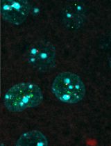
Living cells transfected with the fluorescent protein XRCC1-GFP are micro-irradiated under a confocal microscope and the kinetics of recruitment of the fluorescent protein is followed during 1 min. In our protocol the 405 nm laser is used to induce SSB.
Materials and Reagents
- Glass bottom 35 mm Petri dishes (MatTek, catalog number: P35G-1.5-20-C )
- HeLa adherent cells in culture
- Plasmids coding for the fluorescent protein XRCC1-GFP
Note: The XRCC1 sequence (NCBI P18887) was cloned in the Clontech pEGFP-N1 Vector at the restriction sites EcoRI/ApaI. - Transfection agents (LipoFectamine 2000) (Life Technologies, catalog number: 11668027 )
Note: Currently, it is “Thermo Fisher Scientific, Invitrogen™, catalog number: 11668027”. - DMEM [DMEM(1x) + GlutaMAX™ (Thermo Fisher Scientific, Gibco™, catalog number: 31966-021 )] containing 10% of fetal bovine serum (Thermo Fisher Scientific, Gibco™, catalog number: 16000-044 )
Equipment
- Cell incubator (Thermo Fisher Scientific, model: NAPCO 5400 )
Note: Currently, it is “BioSurplus, catalog number: NAPCO 5400 ”. - Nikon A1 inverted confocal microscope equipped with an environmental chamber allowing the control of temperature, humidity and gas mixture
Note: The lasers used are the following: 405 nm laser for the micro-irradiation, and a 488 nm Argon-laser to visualize the GFP fluorescence. The objective 63x was used.
Software
- The NIS Software
Note: It was used for quantification of the fluorescence intensity at the microirradiation region during time, using the Time Measurement function.
Procedure
文章信息
版权信息
© 2016 The Authors; exclusive licensee Bio-protocol LLC.
如何引用
Campalans, A. and Radicella, J. P. (2016). Laser Microirradiation and Temporal Analysis of XRCC1 Recruitment to Single-strand DNA Breaks. Bio-protocol 6(5): e1746. DOI: 10.21769/BioProtoc.1746.
分类
癌症生物学 > 通用技术 > 遗传学 > DNA损伤
细胞生物学 > 细胞成像 > 共聚焦显微镜
分子生物学 > DNA > DNA 损伤和修复
您对这篇实验方法有问题吗?
在此处发布您的问题,我们将邀请本文作者来回答。同时,我们会将您的问题发布到Bio-protocol Exchange,以便寻求社区成员的帮助。
Share
Bluesky
X
Copy link


