

Immunofluorescent Staining of Mouse Intestinal Stem Cells
小鼠小肠干细胞的免疫荧光染色

发布: 2016年02月20日第6卷第4期 DOI: 10.21769/BioProtoc.1732 浏览次数: 35002
评审: Xuecai GeAnonymous reviewer(s)
参见作者原研究论文
The authors used this protocol in:
Jun 2015


Abstract
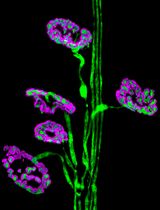
Immunofluorescent staining of organoids can be performed to visualize molecular markers of cell behavior. For example, cell proliferation marked by incorporation of nucleotide (EdU), or to observe markers of intestinal differentiation including paneth cells, goblet cells, or enterocytes (see Figure 1). In this protocol we detail a method to fix, permeabilize, stain and mount intestinal organoids for analysis by immunofluorescent confocal microscopy.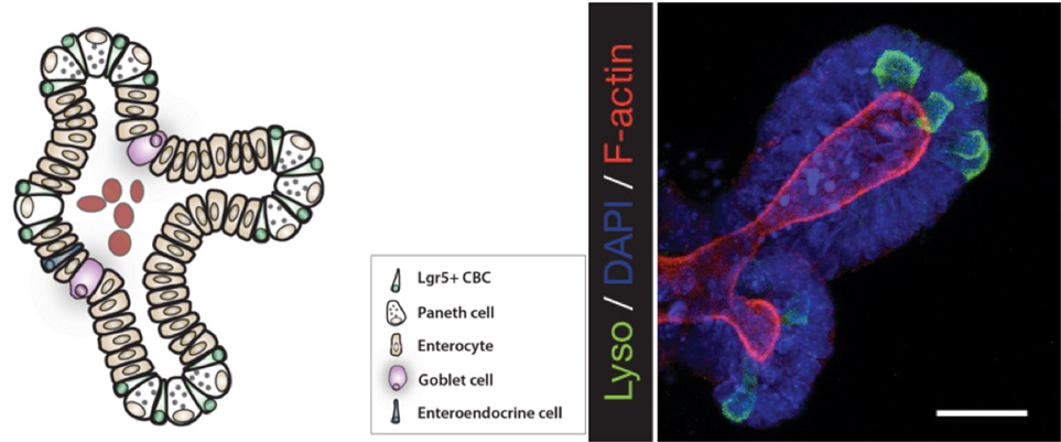
Figure 1. A schematic depicting a crypt-villus forming organoid, and visualization of Paneth cells by immunofluorescence staining. Left: Small intestinal organoids grow as crypt-villus structures that contain all of the multiple differentiated lineages of the intestine. Right: Immunofluorescent staining can be used to visualize individual cell types in the organoid. Here paneth cells are visualized by staining for lysozyme (“Lyso,” Green), which reveals Paneth cells located at crypt bases. F-Actin (Red) reveals crypt structure at the apical surface of the epithelium, and DAPI (Blue) reveals cell nuclei. Scale bar is 25 μm.
Materials and Reagents
- 8 well chamber slides (Thermo Fisher Scientific, Lab-TekTM, catalog number: 154532 )
- Cover Glass, Rectangular #1 (24 x 50 mm, 0.12-0.16 mm) (Corning, catalog number: 2975-245 )
- Primary antibodies
- Rabbit anti-KRT20 (1:200) (Cell Signaling Technology, catalog number: 13063 )
- Rabbit anti-Lysozyme (1:200) (Dako, catalog number: EC 3.2.1.17 )
- Rabbit anti-Muc2 (1:200)/ VHL Antibody (M-20) (Santa Cruz Biotechnology, catalog number: H-300, sc-1534 )
- Villin Antibody (C-19) (1:200) (Santa Cruz Biotechnology, catalog number: sc-7672 )
- Rabbit anti-KRT20 (1:200) (Cell Signaling Technology, catalog number: 13063 )
- Secondary antibodies:
- Goat Anti-rabbit 568 (1:500) (Thermo Fisher Scientific, Molecular Probes, catalog number: 11036 )
Note: Currently, it is “Thermo Fisher Scientific, NovexTM, catalog number: 11036”.
- Donkey Anti-goat 594 (1:500) (Thermo Fisher Scientific, Molecular Probes, catalog number: 11058 )
Note: Currently, it is “Thermo Fisher Scientific, NovexTM, catalog number: 11058”.
- Goat Anti-rat 488 (1:500) (Thermo Fisher Scientific, Molecular Probes, catalog number: a11006 )
Note: Currently, it is “Thermo Fisher Scientific, NovexTM, catalog number: a11006”.
- Goat Anti-rabbit 568 (1:500) (Thermo Fisher Scientific, Molecular Probes, catalog number: 11036 )
- Phosphate Buffered Saline (PBS) (Thermo Fisher Scientific, InvitrogenTM, catalog number: 10010023 )
Note: Currently, it is “Thermo Fisher Scientific, GibcoTM, catalog number: 10010023”.
- Paraformaldehyde (PFA) 16% Solution, EM Grade (Electron Microscopy Sciences, catalog number: 15710-S )
- Freshly prepared 4% PFA in 1x PME Buffer
- ProLong Gold Antifade Mountant (Thermo Fisher Scientific, Molecular Probes, catalog number: P10144 )
Note: Currently, it is “Thermo Fisher Scientific, ProLong®, catalog number: P10144”.
- Clear Nail Polish (available at local drug store)
- Optional
- Click-iT EdU Alexa Fluor 647 for Cell Proliferation (Thermo Fisher Scientific, Invitrogen, catalog number: C10340 )
Note: Currently, it is “Thermo Fisher Scientific, Molecular ProbesTM, catalog number: C10340”.
- Alexa Fluor 647 Phalloidin (Thermo Fisher Scientific, Molecular ProbeTM, catalog number: A22287 )
- BCIP/NBT Substrate Kit (Vector Laboratories, catalog number: SK-5400 )
- 1 μg/ml DAPI for nucleic acid staining (Sigma-Aldrich, catalog number: D9542 )
Note: Materials and equipment to grow organoids prior to fixation (see Isolation, culture, and maintenance of mouse intestinal stem cells)
- Click-iT EdU Alexa Fluor 647 for Cell Proliferation (Thermo Fisher Scientific, Invitrogen, catalog number: C10340 )
- Tris(hydroxymethyl)aminomethane (Sigma-Aldrich, catalog number: 252859 )
- Sodium chloride (NaCl) (Sigma-Aldrich, catalog number: S9888 )
- PIPES (Sigma-Aldrich, catalog number: P6757 )
- Magnesium chloride (MgCl2) (Sigma-Aldrich, catalog number: M8266 )
- Ethylenediaminetetraacetic acid (EDTA) (Sigma-Aldrich, catalog number: E9884 )
- TritonTM X-100 (Sigma-Aldrich, catalog number: X100 )
- TWEEN® 20 (Sigma-Aldrich, catalog number: P2287 )
- Bovine serum albumin (BSA) (Sigma-Aldrich, catalog number: A2058 )
- Tris buffered saline (TBS) (see Recipes)
- 10x PME buffer (see Recipes)
- IF buffer (see Recipes)
- Blocking solution (see Recipes)
- Permeabilization solution (see Recipes)
Equipment
- Benchtop Multi-Purpose Rotator (Thermo Fisher Scientific, model: 2309 )
- Leica Inverted Confocal SP8 equipped with a White Light Laser, a Leica HyD Detector and the Leica Application Suite software
Software
- Leica Application Suite
Procedure
文章信息
版权信息
© 2016 The Authors; exclusive licensee Bio-protocol LLC.
如何引用
O’Rourke, K. P., Dow, L. E. and Lowe, S. W. (2016). Immunofluorescent Staining of Mouse Intestinal Stem Cells. Bio-protocol 6(4): e1732. DOI: 10.21769/BioProtoc.1732.
分类
干细胞 > 成体干细胞 > 肠道干细胞
细胞生物学 > 组织分析 > 组织染色
您对这篇实验方法有问题吗?
在此处发布您的问题,我们将邀请本文作者来回答。同时,我们会将您的问题发布到Bio-protocol Exchange,以便寻求社区成员的帮助。