

Single Molecule RNA FISH in the Mammalian Oocyte
哺乳动物卵母细胞中的单分子RNA荧光原位杂交(FISH)

发布: 2015年12月05日第5卷第23期 DOI: 10.21769/BioProtoc.1666 浏览次数: 11286
评审: Anonymous reviewer(s)
参见作者原研究论文
The authors used this protocol in:

Jan 2015


Abstract
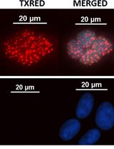
RNA fluorescence in situ hybridization is a method to localize and measure gene expression in individual cell or tissue. Using multiple specific fluorescently labeled oligonucleotides greatly increases signal-to-noise ratio and thus enables detection of single RNA molecule. Around forty different DNA oligonucleotides designed to common RNA target and labeled with single fluorophore at 3´ terminus hybridizes with target RNA in fixed cells. We adapt this method to visualize target RNA in the mammalian oocyte. The ability to detect single transcript in the mammalian oocyte was challenging due to its large cell size. This method consists of four simple steps: fixation, permeabilization, hybridization and imaging. The protocol is adapted to this large nonattached cell to visualize maternal RNAs.
Combination of various fluorophores allows detection of more RNA targets. This method might be used with organelle markers or expanded with immunofluorescence protocol.
Materials and Reagents
- Cover glasses thickness No.1 22 x 22 mm (Marienfeld-Superior, catalog number: 0101050 )
- Fisherfinest Premium Microscope Slides (Thermo Fisher Scientific, catalog number: 125447 )
- Tissue culture 24-well plates (TPP Techno Plastic Products AG, catalog number: 92024 )
- Tissue culture 96-well plates (TPP Techno Plastic Products AG, catalog number: 92096 )
- Borosilicate glass capillary 3.3. (Hilgenberg GmbH, catalog number: 1409363 ), hand pipette for manipulation) with tip diameter around 100 µm (Hilgenberg GmbH)
- Microtube from Eppendorf 15 ml RNase free (Sigma-Aldrich, catalog number: 0030123.328 )
Note: Pricing & availability is not currently available. - Aluminum foil (29 cm x 150 m) (MAIS.s.r.o.)
- Oocytes from Six-weeks-old female CD1 mice; isolation of mouse oocytes described in Bio-protocol (Tetkova and Hancova, 2015), also (Susor et al., 2015)
- Paraformaldehyde final concentration 4% in PBS (store at -20 °C) (Thermo Fisher Scientific, Afla Aesar, catalog number: 30525894 )
Note: Currently, it is “Sigma-Aldrich, catalog number: 30525894 ”. - Triton X-100 final concentration 2% in 1x PBS (fresh) (Sigma-Aldrich, catalog number: 9002931 )
- 20x saline-sodium citrate (SSC) stock is diluted in RNase free water, in the same day of experiment (Sigma-Aldrich, catalog number: S6639 )
- Ethanol 100% (for final concentration 70% ethanol, freshly dilute 100% ethanol with DEPC water) (Merck Millipore Corporation, catalog number: 1085430250 )
- VECTASHIELD HardSetTM with DAPI (storage 2-8 °C) (Vector Laboratories, catalog number: H1500 )
- ProtectRNA RNase Inhibitor 500x concentrated (final working concentration is 1x; storage at 2-8 °C in dark) (Sigma-Aldrich, catalog number: R7397 )
- Stellaris probes (stock concentration 5 nmol) (Bioresearch Technologies)
- Nuclease-free water (Life Technologies, Ambion®, catalog number: AM9932 )
Note: Currently, it is “Thermo Fisher Scientific, AmbionTM, catalog number: AM9932”. - Phosphate buffered saline (PBS) (Sigma-Aldrich, catalog number: P4417 )
- Poly(vinyl alcohol) (PVA) (Sigma-Aldrich, catalog number: 341584 )
- Formamide (5 ml for 10% final concentration) (Sigma-Aldrich, catalog number: F7503 )
- Dextran sulfate sodium salt from Leuconostoc spp. (Sigma-Aldrich, catalog number: D8906 )
- Wash buffer (see Recipes)
- Hybridization buffer (see Recipes)
- Isolation buffer PVA (see Recipes)
Equipment
- Stereomicroscope Zeiss 2000C (Thermo Fisher Scientific)
- Heating plate (P-lab)
- Multi Bio 3D mini-Shaker (Biosan Laboratories)
- Thermostat incubator mini I5110 (Labnet International)
- Confocal microscope Leica SP5 (Leica Microsystems)
- Filter sets appropriate for fluorophores and excitation lasers
- 40x or 63x or 100x oil immersion objective
Procedure
文章信息
版权信息
© 2015 The Authors; exclusive licensee Bio-protocol LLC.
如何引用
Jansova, D. (2015). Single Molecule RNA FISH in the Mammalian Oocyte. Bio-protocol 5(23): e1666. DOI: 10.21769/BioProtoc.1666.
分类
细胞生物学 > 细胞染色 > 核酸
您对这篇实验方法有问题吗?
在此处发布您的问题,我们将邀请本文作者来回答。同时,我们会将您的问题发布到Bio-protocol Exchange,以便寻求社区成员的帮助。