

Metabolic Assays for Detection of Neutral Fat Stores
代谢试验检测中性脂肪储存
发布: 2015年06月20日第5卷第12期 DOI: 10.21769/BioProtoc.1511 浏览次数: 15693
评审: HongLok LungJustine MarsolierKate Hannan
参见作者原研究论文
The authors used this protocol in:
Dec 2013
Advertisement


Abstract
Lipid droplets (LDs) are ubiquitous intracellular structures whose formation, growth, and maintenance are highly regulated (Wang et al., 2013; Ranall et al., 2011; Goodman, 2009). Lipid metabolism and droplet dynamics are of considerable interest to agriculture, biofuel production, viral pathology, nutrition, and cancer biology (Walther and Farese, 2009; Liu et al., 2010). Accumulation of fatty acids and neutral lipids in nonadipose tissues is cytotoxic (Kourtidis et al., 2009). BODIPY 493/503 (4,4-Difluoro-1,3,5,7,8-Pentamethyl-4-Bora-3a,4a-Diaza-s-Indacene) is the standard dye to study LDs within adipocytes. BODIPY 493/503 contains a nonpolar structure that, upon binding to neutral lipid, emits a green fluorescence signal with a narrow wavelength range, making it an ideal fluorophore for multi-labeling experiments. The hydrophobic nature of the dye molecules promotes rapid entry into the nonpolar environment of LDs (Listenberge and Brown, 2007). Gocze and Freeman showed that the lipid fluorescent variability is significantly lower when using BODIPY493/503 compared to Nile Red, suggesting that it may be more specific for the LD (Gocze and Freeman, 1994). Here, we describe a BODIPY 493/503 assay for the detection of neural fat stores in cultured cells (Figure 1) (Wang et al., 2013).
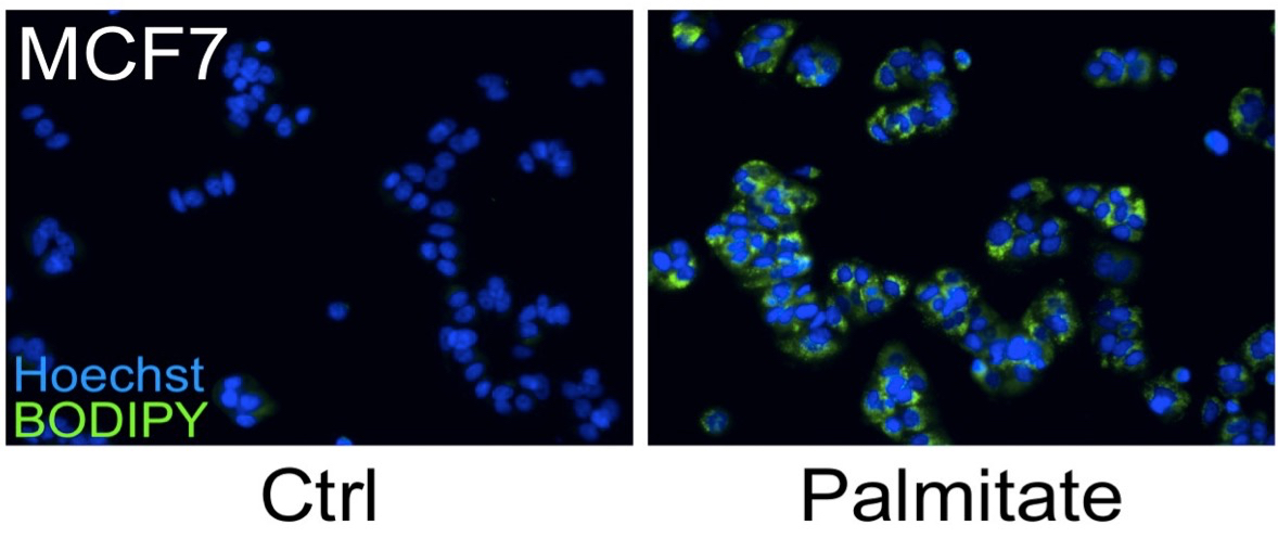
Figure 1. MCF7 cells were treated with 250 μM palmitate or vehicle control for 24 h. A number of breast cancer cells possess a lipogenic metabolic phenotype that makes them especially sensitive to the addition of physiological concentrations of exogenous saturated fatty acids, such as palmitate. Although palmitate supplementation induces cell death in HER2/neu-positive cells, other breast cancer sub-types, including MCF-7 cells, accumulate the fatty acid which leads to significant increases in intracellular triglyceride fat stores. Cells were fixed and stained for fat stores with BODIPY 493/503 (green). Hoechst 33342 (blue) was used for nuclei staining.
Materials and Reagents
- Cell line suitable for testing, MCF7 (ATCC, catalog number: HTB-22 TM) cells perform well as a staining control
- Dulbecco’s Modified Eagle’s Medium (DMEM) (high glucose with L-glutamine) (Thermo Fisher Scientific, catalog number: SH3024301 ) or other media appropriate for MCF7 cell culture
- Fetal bovine serum (FBS) (Sigma-Aldrich, catalog number: F4135 )
- Phosphate buffered saline (PBS) (e.g. HyClone, catalog number: SH30258-02 ) or Dulbecco's phosphate buffered saline (with Ca2+ and Mg2+) (DPBS) (e.g. Sigma-Aldrich, catalog number: D1283 )
- Sodium palmitate, (used as positive control) (Sigma-Aldrich, catalog number: P9767 )
- Dimethyl sulfoxide (Sigma-Aldrich, catalog number: D8418 )
- BODIPY 493/503 (4,4-Difluoro-1,3,5,7,8-Pentamethyl-4-Bora-3a,4a-Diaza-s-Indacene) (Life Technologies, catalog number: D-3922 )
- 37% formaldehyde (Thermo Fisher Scientific, catalog number: F79 )
- Hoechst 33342 (Life Technologies, catalog number: H21492 )
- 500x BODIPY 493/503 stock solution (see Recipes)
- 10,000x Hoechst 33342 stock solution (see Recipes)
Equipment
- 96-well tissue culture plate suitable for imaging, (e.g. Corning, Costar®, catalog number: 3603 )
- Cell culture incubator at 37 °C with 5% CO2
- Fluorescence microscope or automated imaging system (e.g. IN Cell Analyzer, GE Healthcare)
Procedure
文章信息
版权信息
© 2015 The Authors; exclusive licensee Bio-protocol LLC.
如何引用
Baumann, J. M., Kokabee, L., Wang, X., Sun, Y., Wong, J. and Conklin, D. S. (2015). Metabolic Assays for Detection of Neutral Fat Stores. Bio-protocol 5(12): e1511. DOI: 10.21769/BioProtoc.1511.
分类
癌症生物学 > 通用技术 > 细胞生物学试验 > 新陈代谢
生物化学 > 脂质 > 脂质测定
细胞生物学 > 细胞染色 > 脂质
您对这篇实验方法有问题吗?
在此处发布您的问题,我们将邀请本文作者来回答。同时,我们会将您的问题发布到Bio-protocol Exchange,以便寻求社区成员的帮助。