

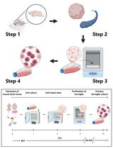
Murine Liver Myeloid Cell Isolation Protocol
鼠肝髓样细胞分离法

(*contributed equally to this work) 发布: 2015年05月20日第5卷第10期 DOI: 10.21769/BioProtoc.1471 浏览次数: 33165
评审: Fanglian HeAnonymous reviewer(s)
参见作者原研究论文
The authors used this protocol in:
Sep 2014
Advertisement


Abstract
In homeostasis, the liver is critical for the metabolism of nutrients including sugars, lipids, proteins and iron, for the clearance of toxins, and to induce immune tolerance to gut-derived antigens. These functions predispose the liver to infection by blood-borne pathogens, and to a variety of diseases ranging from toxin and medication-induced disorders (CCl4, acetaminophen) to metabolic disorders (steatohepatitis, alcoholic liver disease, biliary obstruction, cholestasis) or autoimmunity. Chronic liver injury often progresses to life threatening fibrosis and can end in liver cirrhosis and hepatocellular carcinoma (Pellicoro et al., 2014).
The liver contains parenchymal cells or hepatocytes that make up the majority of hepatic cells. It also contains non-parenchymal structural cells such as sinusoidal endothelial cells and a large number of non-parenchymal innate immune cells, mainly monocytes, neutrophils, macrophages, DCs, NK and NKT cells that can trigger an adaptive immune response in the case of infections or other pathogenic insults (Jenne and Kubes, 2013). How this immune response is regulated determines the extent of acute and chronic liver injury (Stijlemans et al., 2014). In this context, liver macrophages have been demonstrated to play central but divergent (from initiating to resolving) functions in liver injury (Sica et al., 2014). It has become clear in the last years that hepatic macrophages consist of two classes, tissue-resident macrophages, the Kupffer cells (KCs) originating from yolk sac/fetal liver progenitors and tissue-infiltrating macrophages originating from bone marrow-derived Ly6CHi monocytes (Jinhoux and Jung, 2014; Tacke and Zimmerman, 2014). Distinguishing the activities of KCs from those of monocyte-derived macrophages during liver injury or repair is currently a frontline research topic in the macrophage field. Indeed, considering that clinical management of liver failure remains problematic, a better understanding of the immune mechanisms regulating liver injury is expected to allow the development of new therapeutic modalities. Here, we describe an isolation technique for liver non-parenchymal polymorphonuclear (PMN) and mononuclear myeloid cells permitting their molecular and functional characterization.
Materials and Reagents
- 7-8 weeks old female C57Black/6 mice (Janvier Labs)
- RPMI-1640 medium (RPMI) (Life Technologies, catalog number: 52400-041 )
- Collagenase Type III (Worthington Biochemical, catalog number: LS004180 )
- DNase I (Roche Diagnostics, catalog number: 04536282001 )
- Heparin (sodium salt from porcine intestinal mucosa) (Sigma-Aldrich, catalog number H3393-1MU )
- Hank’s buffered salt solution (HBSS) without calcium or magnesium or phenol red (Life Technologies, Gibco®, catalog number: 14175-053 )
- NH4Cl (Merck KGaA, catalog number: 01145.0500 )
- KHCO3 (Merck KGaA, catalog number: 04854.0500 )
- EDTA (Duchefa Biochemie, catalog number: E0511.1000 )
- HCl (37% stock solution) (Merck KGaA, catalog number: 1.00317.1000 )
- Fetal bovine serum (FBS) (BiowhittakerTM/Lonza, catalog number: DE14-801F )
- LymphoprepTM (Axis-shield, catalog number: 1114547 )
- PercollTM (GE Healthcare, catalog number: 17-0891-01 )
- Purified CD16/CD32 (Fc-Block) (clone 2.4G2) (BD Biosciences, catalog number: 553142 )
- PE-Cy7-conjugated anti-CD11b antibody (clone M1/70) (BD Biosciences, catalog number: 552850 )
- AF647-conjugated anti-Ly6C antibody (clone ER-MP20) (Serotec, catalog number: MCA2389A647 )
- PerCP-Cy5.5-conjugated anti-I-A/I-E (MHC-II) antibody (clone M5/114.15.2) (Biolegend, catalog number: 107626 )
- FITC-conjugated anti-Ly6G antibody (clone 1A8) (BD Biosciences, catalog number: 551460 )
- APC-Cy7-conjugated CD45 antibody (clone 30-F11) (BD Biosciences, catalog number: 103116 )
- PE-conjugated F4/80 antibody (clone CI: A3-1) (AbD Serotec, catalog number: MCA497PET )
- Trypan blue (BDH Chemicals, catalog number: 34078 )
- NaCl (Thermo Fisher Scientific, catalog number: 10428420 )
- KH2PO4 (Merck KGaA, catalog number: 1.04873.1000 )
- Na2HPO4.2H2O (Merck KGaA, catalog number: 1.06580.1000 )
- L-glutamine (Sigma-Aldrich, catalog number: G8540-100G )
- Penicillin (Life Technologies, Gibco®, catalog number: 15140-122 )
- Streptomycin (Life Technologies, Gibco®, catalog number: 15140-122)
- β-mercaptoethanol (Sigma-Aldrich, catalog number: M3148 )
- Sodium pyruvate (Life Technologies, Gibco®, catalog number: 11360-039 )
- Non-essential amino acids (Life Technologies, Gibco®, catalog number: 11140-035 )
- Liver digestion medium (see Recipes)
- Phosphate buffered saline (PBS) (see Recipes)
- 33% Percoll working solution (see Recipes)
- Erythrocyte lysis buffer (see Recipes)
- MACS buffer (see Recipes)
- Cell suspension medium (see Recipes)
- Blocking medium (see Recipes)
- Complete medium (see Recipes)
- Trypan blue working solution (see Recipes)
Equipment
- Polyester filters cut in 10 x 10 cm squares, thread diameter 70 μm (Spectrumlabs, catalog number: 146490 )
- 10 ml syringes (Omnifix, catalog number: 473203 )
- BD Falcon 50 ml polypropylene tubes (BD Biosciences, catalog number: 2070 )
- BD Falcon 15 ml polypropylene tubes (BD Biosciences, catalog number: 2096 )
- BD Falcon 5 ml polypropylene round-bottom tube (BD Biosciences, catalog number: 352063 )
- Needles (Microlance 22G1 ½, 0.7 * 40 mm) (BD Biosciences, ref: 301000 )
- Sterile culture hood
- Surgical scissors and forceps
- 37°C, 5% CO2 cell culture incubator (Forma Scientific)
- Pipettes
- Centrifuges (Eppendorf, models: 5810R and 5417C )
- Orbital shaker (Belgolabo, model: Julabo type SW-20C ) used at 200 rpm
- Light microscope (Olympus, model: CK2 )
- Multicolor flow cytometer (BD Biosciences, FACSCantoTM )
- GentleMACSTM Dissociator (Miltenyi Biotec, catalog number: 130-093-235 )
- GentleMACSTM C-tubes (Miltenyi Biotec, catalog number: 130-093-237 )
Procedure
文章信息
版权信息
© 2015 The Authors; exclusive licensee Bio-protocol LLC.
如何引用
Stijlemans, B., Sparkes, A., Abels, C., Keirsse, J., Brys, L., Elkrim, Y., Baetselier, P. D., Beschin, A. and Ginderachter, J. A. V. (2015). Murine Liver Myeloid Cell Isolation Protocol . Bio-protocol 5(10): e1471. DOI: 10.21769/BioProtoc.1471.
分类
免疫学 > 免疫细胞分离 > 骨髓细胞
细胞生物学 > 细胞分离和培养 > 细胞分离
您对这篇实验方法有问题吗?
在此处发布您的问题,我们将邀请本文作者来回答。同时,我们会将您的问题发布到Bio-protocol Exchange,以便寻求社区成员的帮助。