

Histochemical Detection of Zn in Plant Tissues
植物组织中锌的组织化学检测


(*contributed equally to this work) 发布: 2015年05月20日第5卷第10期 DOI: 10.21769/BioProtoc.1470 浏览次数: 11494
评审: Maria SinetovaAnonymous reviewer(s)
参见作者原研究论文
The authors used this protocol in:
Jul 2014


Abstract
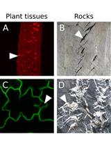
Accumulation of metals in plant tissues, and occasionally, different cells of the same tissue, may be highly non-uniform (Seregin and Kozhevnikova, 2008). Easy-to-use histochemical methods may greatly help to investigate the distribution and accumulation of metals within and among plant tissues, and also provide information on their subcellular localization (Seregin and Kozhevnikova, 2011). The histochemical techniques of zinc (Zn) visualization are based on the formation of the blue-colored complex of Zn with the metallochrome indicator Zincon (C20H15N4NaO6S), or the green-fluorescent complex with Zinpyr-1 (C46H36Cl2N6O5) (Seregin et al., 2011; Seregin and Kozhevnikova, 2011). A method for histochemical Zn detection in plant tissues using Zinpyr-1 was first proposed by Sinclair et al. (2007), and later modified by Seregin et al. (2011), and Seregin and Kozhevnikova (2011). Histochemical data supplement the results of quantitative analysis, thus allowing a detailed study of the distribution, accumulation, and translocation pathways of Zn within the plant, which are important topics in modern plant physiology. These histochemical techniques have been successfully applied in different plant species, for example Zea mays (Seregin et al., 2011), Noccaea caerulescens and Thlaspi arvense (Kozhevnikova et al., 2014a), Capsella bursa-pastoris and Lepidium ruderale (Kozhevnikova et al., 2014b), in which Zn was detected in different root and shoot tissues. Here, we present the full staining protocols for these methods, developed or modified in our lab (Seregin and Kozhevnikova, 2011; Kozhevnikova et al., 2014a; Kozhevnikova et al., 2014b).
Keywords: Zinc localization (锌的定位)Materials and Reagents
- Staining with Zinpyr-1 (see Recipes)
- Zinpyr-1 or 4′, 5′-Bis[bis(2-pyridylmethyl)aminomethyl]-2′,7′-dichlorofluorescein (C46H36Cl2N6O5, Mr= 823.72) (Sigma-Aldrich, catalog number: 40667 or Millitech, catalog number: ZP1 )
- Dimethyl sulfoxide or DMSO (C2H6OS)
- Ethylenediaminetetraacetic acid (disodium salt) (Na2EDTA, C10H14O8N2Na2⋅2H2O, Mr = 372.24)
- Super demineralized water
- Zinpyr-1 or 4′, 5′-Bis[bis(2-pyridylmethyl)aminomethyl]-2′,7′-dichlorofluorescein (C46H36Cl2N6O5, Mr= 823.72) (Sigma-Aldrich, catalog number: 40667 or Millitech, catalog number: ZP1 )
- Staining with Zincon (see Recipes)
- Zincon sodium salt (C20H15N4NaO6S, Mr = 462.4) (Sigma-Aldrich, catalog number: 201332 )
- Borax (Na2B4O7⋅10 H2O)
- Sodium hydroxide (NaOH)
- Ethylenediaminetetraacetic acid, disodium salt (Na2EDTA, C10H14O8N2Na2⋅2H2O, Mr = 372.24)
- Super demineralized water
- Zincon sodium salt (C20H15N4NaO6S, Mr = 462.4) (Sigma-Aldrich, catalog number: 201332 )
Equipment
- Light microscope with a color digital camera attachment (staining with Zincon); confocal microscope or fluorescence microscope with appropriate filters and digital camera attachment (staining with Zinpyr-1; see below for spectral characteristics of the dye)
- Micro pipettes (100-1,000 µl) and pipette tips
- Vortex
- Precision balances
- Razor blades
- 50 ml, 100 ml and 1 L flasks
- 2 ml microtubes
- 5 ml or 15 ml centrifuge tubes
- Heat-resistant 20 ml flask
- Microscope slides and cover glasses
- Tweezers
- Dissecting needles
- Magnetic stirrer with heating
- Axio Imager Z2 microscope (ZEISS)
Procedure
文章信息
版权信息
© 2015 The Authors; exclusive licensee Bio-protocol LLC.
如何引用
Seregin, I., Kozhevnikova, A. and Schat, H. (2015). Histochemical Detection of Zn in Plant Tissues. Bio-protocol 5(10): e1470. DOI: 10.21769/BioProtoc.1470.
分类
植物科学 > 植物细胞生物学 > 组织分析
植物科学 > 植物生理学 > 离子分析
细胞生物学 > 细胞染色 > 其它化合物
您对这篇实验方法有问题吗?
在此处发布您的问题,我们将邀请本文作者来回答。同时,我们会将您的问题发布到Bio-protocol Exchange,以便寻求社区成员的帮助。