

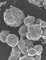
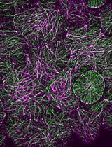
Imaging and Quantitative Analysis of Size and Distribution of Spherical Bodies, e.g. Embryonic Oil Bodies
胚胎油体等球状体大小、分布的成像和定量测定


发布: 2015年01月05日第5卷第1期 DOI: 10.21769/BioProtoc.1369 浏览次数: 9076
评审: Renate WeizbauerArsalan DaudiAnonymous reviewer(s)
参见作者原研究论文
The authors used this protocol in:
Apr 2014
Advertisement


Abstract
Oil bodies (OBs) are seed-specific lipid storage organelles that allow the accumulation of neutral lipids that sustain plantlet development after the onset of germination. Using fluorescent dyes and confocal microscopy, we monitored the dynamics of OBs in living Arabidopsis (Arabidopsis thaliana) embryos at different stages of development (Miquel et al., 2014). Image acquisition was followed by a detailed statistical analysis of OB size and distribution during seed development in the four dimensions (x, y, z, and t).
Keywords: Oil body (油体)Materials and Reagents
- Plant material
- Arabidopsis thaliana plants, wild type or mutant or transgenic plants.
- Developing siliques between 6 and 11 days after pollination of plants grown in a greenhouse under the following conditions (13 h of light, diurnal temperature of 25 °C, and nocturnal temperature of 17 °C), and irrigated twice per week with mineral nutrient solution.
- Arabidopsis thaliana plants, wild type or mutant or transgenic plants.
- Nile Red (a neutral lipid stain, at 2 μg/ml final in acetone) (Sigma-Aldrich, catalog number: 72485 )
Equipment
- Confocal microscope
Note: In this study an inverted LEICA SP2-AOBS spectral confocal laser microscope (Leica Microsystems) equipped with an HCX PL APO CS 40 x 1.25 objective and a multiline argon laser was used. - Forceps (Dumont No.5) (Sigma-Aldrich)
- Scalpel (11 P, blade) (Swann Norton)
- Glass slides or glass-bottom dish, cover slips
- Binocular (Nikon Corporation, model: SMZ1000 ) (Champigny sur Marne with a led ring - Shott easyledTM for illumination)
Software
- ND-SAFIR (http://serpico.rennes.inria.fr/doku.php?id=software:nd-safir:index)
- AVIZO® FIRE (http://www.vsg3d.com/avizo/fire)
Procedure
文章信息
版权信息
© 2015 The Authors; exclusive licensee Bio-protocol LLC.
如何引用
Readers should cite both the Bio-protocol article and the original research article where this protocol was used:
- Miquel, M., Trigui, G., Trubuil, A. and Dubreucq, B. (2015). Imaging and Quantitative Analysis of Size and Distribution of Spherical Bodies, e.g. Embryonic Oil Bodies. Bio-protocol 5(1): e1369. DOI: 10.21769/BioProtoc.1369.
- Miquel, M., Trigui, G., d'Andrea, S., Kelemen, Z., Baud, S., Berger, A., Deruyffelaere, C., Trubuil, A., Lepiniec, L. and Dubreucq, B. (2014). Specialization of oleosins in oil body dynamics during seed development in Arabidopsis seeds. Plant Physiol 164(4): 1866-1878.
分类
植物科学 > 植物细胞生物学 > 细胞染色
植物科学 > 植物细胞生物学 > 细胞成像
细胞生物学 > 细胞成像 > 活细胞成像
您对这篇实验方法有问题吗?
在此处发布您的问题,我们将邀请本文作者来回答。同时,我们会将您的问题发布到Bio-protocol Exchange,以便寻求社区成员的帮助。