

Imaging and Measurement of Nanomechanical Properties within Primary Xylem Cell Walls of Broadleaves
阔叶初生木质部细胞壁内的纳米力学性能的成像和测定

发布: 2014年12月20日第4卷第24期 DOI: 10.21769/BioProtoc.1360 浏览次数: 13232
评审: Arsalan DaudiMasahiro MoritaAnonymous reviewer(s)
参见作者原研究论文
The authors used this protocol in:

Jul 2014


Abstract
A technique of atomic force microscopy (AFM) called PeakForce quantitative nanomechanical mapping (PeakForce QNM) is an efficient tool for the quantitative mechanobiological imaging of fibrillar aggregate, human epidermal cell and woody plant cell wall topography (Sweers et al., 2011; Heu et al., 2012; Ďurkovič et al., 2012; Ďurkovič et al., 2013). Here, we describe a detailed protocol for the measurement of nanomechanical properties of primary xylem cell walls in woody plants, for the determination of reduced Young’s modulus of elasticity (MOE), adhesion, deformation, and energy dissipation (Figure 1). This new technique provides direct control of the maximum loading force and the deformation depth in cell wall samples keeping indentations small, while at the same time eliminating damaging lateral forces in order to preserve both the AFM tip and plant sample. High-resolution and non-destructive imaging shed new quantitative mechanistic insights into the structural biology of woody plant cell walls. This procedure can also be adapted for other biological samples with a varying range of stiffness.
Keywords: Atomic force microscopy (原子力显微镜)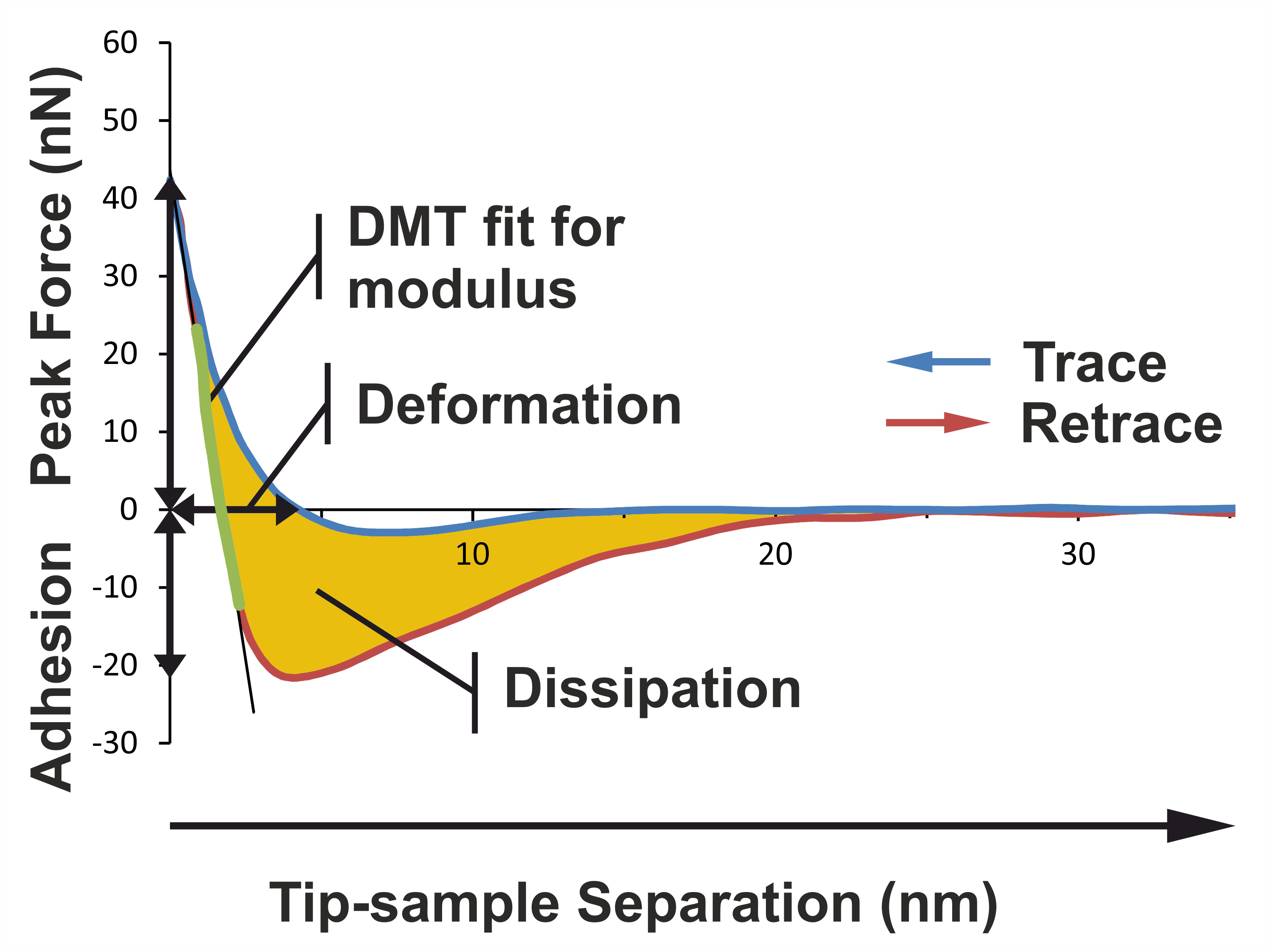
Figure 1. Basic principles illustrating how the different nanomechanical properties are extracted from the force curve. The image shows a typical force curve for a primary xylem cell wall sample in the Dutch elm disease-sensitive hybrid 'Groeneveld'. PeakForce QNM mode performs a very fast force curve at every pixel in the image. Analysis of force curve data is done on the fly, providing a map of multiple nanomechanical properties that has the same resolution as the height image. The adhesion is the vertical distance between the base line and the lowest portion of the retraction curve. The deformation is the horizontal distance between the contact point and the turn-away point (representing maximum indentation). The Young’s modulus of elasticity can be extracted by extrapolating the linear portion of the retraction curve after the contact point using a Derjaguin, Muller, Toporov (DMT) model fit. The slope of the linear portion is determined according to the least square method procedure. The energy dissipation can be calculated by integrating the area between the two curves.
Materials and Reagents
- Leaf midribs from broadleaves (Ulmus spp., Sorbus spp.)
- Glutaraldehyde solution, 25% in H2O (Grade II) (Sigma-Aldrich, catalog number: G6257 )
- Ethanol solutions (Analytical Reagents)
Note: 10%, 20%, 30%, 40%, 50%, 60%, 70%, 80%, 90%, each of them prepared by diluting of 96% ethanol in a 0.1 M cacodylate buffer, and 100% ethanol.
- 25% glutaraldehyde (Grade I) (Sigma-Aldrich, catalog number: G5882 )
- Xylene (Analytical Reagents) or alternative cleaners with a lower toxicity (for example D-limonene)
Note: Work with xylene or D-limonene only in a properly operating and certified fume cupboard.
- Paraplast plus (Sigma-Aldrich, catalog number: P3683 )
- (3-Aminopropyl) triethoxy-silane (Sigma-Aldrich, catalog number: A3648 )
- 0.1 M cacodylate buffer (see Recipes)
- Circle glass slides coated with (3-aminopropyl)triethoxy-silane (see Recipes)
- 1x phosphate buffered saline (PBS) (see Recipes)
- Glutaraldehyde fixative solution (see Recipes)
Equipment
- Razor blades (fine tweezers and heated forceps)
- pH meter
- Exicator with a vacuum pump
- Petri dishes
- Laboratory drying chamber
- Retracting base sledge microtome (Series 8000) (Bright Instrument Co.)
- Circle glass slides (11 mm in diameter, 1 mm thick)
- Slide warmer
- Steel AFM sample mounting disks
- Sample adhesive pads
- Silicon probes MPP-12120 (model: TAP150A ) having a 180° rotated tip and the spring constant at least 5 N/m (Bruker AFM Probes)
- Fused silica reference sample (nominal MOE 72.9 GPa) or any other reference sample having the MOE value higher than 70 GPa
- TGT1 commercial tip characterization grating (NT-MDT Co.)
- MultiMode 8 atomic force microscope with a Nanoscope V controller and a PeakForce Quantitative Nanomechanical Mapping mode (Bruker Corporation)
Software
- NanoScope analysis (version 1.40r2) (Bruker Corporation)
- MATLAB (version 7) (MathWorks)
Procedure
文章信息
版权信息
© 2014 The Authors; exclusive licensee Bio-protocol LLC.
如何引用
Ďurkovič, J., Kardošová, M. and Lagaňa, R. (2014). Imaging and Measurement of Nanomechanical Properties within Primary Xylem Cell Walls of Broadleaves. Bio-protocol 4(24): e1360. DOI: 10.21769/BioProtoc.1360.
分类
植物科学 > 植物细胞生物学 > 细胞成像
细胞生物学 > 细胞结构 > 细胞粘附
细胞生物学 > 细胞成像 > 固定细胞成像
您对这篇实验方法有问题吗?
在此处发布您的问题,我们将邀请本文作者来回答。同时,我们会将您的问题发布到Bio-protocol Exchange,以便寻求社区成员的帮助。