

Combined in situ Hybridization/Immunohistochemistry (ISH/IH) on Free-floating Vibratome Tissue Sections
自由浮动振动切片机中组织切片的组合原位杂交/免疫组织化学法(ISH/IH)

发布: 2014年09月20日第4卷第18期 DOI: 10.21769/BioProtoc.1243 浏览次数: 15489
评审: Hui ZhuXuecai GeAnonymous reviewer(s)
参见作者原研究论文
The authors used this protocol in:

Sep 2012


Abstract
In situ hybridization and immunostaining are common techniques for localizing gene expression, the mRNA and protein respectively, within tissues. Both techniques can be applied to tissue sections to achieve similar goals, but in some cases, it is necessary to use them together. For example, complement C1q is a secreted protein complex that can target the innate immune response during inflammation. Complement has been found to be elevated early and before severe neurodegeneration in several disease models. Thus, complement may serve as an important marker for disease progression and may contribute to the pathology under certain conditions. Since complement is a secreted complex, immunostaining for C1q does not necessarily reveal where compliment is produced. In situ hybridization for complement components, C1q a, b, or c mRNA, is ideal to mark complement producing cells in tissue. In situ hybridization can be coupled with cell-type-specific immunostaining for accurate identification of the cell types involved. Protein localization and mRNA localization together can reveal details as to the relationship between complement producing and complement target cells within disease tissues. Here we outline the steps for combined in situ hybridization and immunostaining on the same tissue section. The protocol outlined here has been designed for detection of complement C1q in neurons and microglia in the mouse brain.
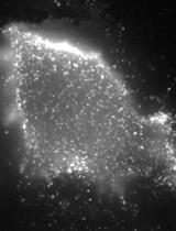
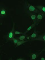
Provided here are two approaches for combined ISH/IH. In the 1st example, in situ hybridization of C1q mRNA is performed together with fluorescent detection of Purkinje neuron cell bodies using Calbindin-D28K antibody. In the 2nd example, C1q mRNA in situ is performed together with 3,3’-diaminobenzidine (DAB) detection of microglia using CD68 antibody. Please note that modifications to the protocol may be needed for the use of distinct probes and antibodies, as well as alternate tissue-processing methods that are not specified herein. For appropriate examples of procedure results, please see images published in Lopez et al.. (2012).
Materials and Reagents
- Mice (to be obtained from appropriate sources and in accordance with approved animal regulations)
- >200 ng/ml of purified DIG-labeled RNA probe [See Stevens et al. (2007) for source of C1q RNA probe. In general, size of probe can range from 500-1,500 bp or more. Probe size alone has not appeared to affect tissue penetration or alter background. Larger probes may have enhanced signal detection.]
- RNaseZAP (Life Technologies, catalog number: AM9780 )
- RNase free water (Life Technologies, catalog number: 10977-023 )
- 32% paraformaldehyde solution (VWR International, catalog number: 100496-496 )
- 10x RNase free phosphate buffer saline (PBS) (Life Technologies, catalog number: AM9624 )
- 20x RNase-free SSC buffer (Life Technologies, catalog number: 15557-036 )
- Formamide (Sigma-Aldrich, catalog number: 47670 )
- Tween-20 (Sigma-Aldrich, catalog number: P1379 )
- tRNA (Roche Diagnostics, catalog number: 10109525001 )
- Salmon testis DNA (Sigma-Aldrich, catalog number: D9156 )
- Heparin salt (Sigma-Aldrich, catalog number: H4784 )
- Sodium dodecyl sulfate (SDS) (Sigma-Aldrich, catalog number: L3771 )
- Bovine serum albumin (BSA) (Sigma-Aldrich, catalog number: A9647 )
- Triton X-100 (Sigma-Aldrich, catalog number: X100 )
- Anti-Digoxigenin-AP (fab fragment) (Roche Diagnostics, catalog number: 11093274910 )
- NBT/BCIP ready-to-use (Roche Diagnostics, catalog number: 11697471001 )
- 30% hydrogen peroxide
- Methanol
- Diethyl pyrocarbonate (DEPC) (Sigma-Aldrich, catalog number: D5758 )
- Rat Anti-CD68 (Bio-Rad Laboratories, AbD Serotec, catalog number: MCA1957GA )
- Rabbit Anti-Calbindin-D28k (Sigma-Aldrich, catalog number: C2724 )
- Fluoromount-G (Thermo Fisher Scientific, catalog number: OB100-01 )
- Anti-Rabbit Alexa 488 (Life Technologies, catalog number: A21206 )
- Anti-Rat HRP (Jackson ImmunoResearch Laboratories, catalog number: 712035150 )
- SIGMA FAST DAB (Sigma-Aldrich, catalog number: D4293 )
- HNPP/Fast Red TR (Roche Diagnostics, catalog number: 11758888001 )
- NBT/BCIP tablet (Roche Diagnostics)
- PFA (see Recipes)
- 5x SSC (see Recipes)
- Hybe (see Recipes)
- Wash 1 (see Recipes)
- Wash 2 (see Recipes)
- Block (see Recipes)
- DEPC block (see Recipes)
- HPM (see Recipes)
- Detection buffer (see Recipes)
- Stop buffer (see Recipes)
Equipment
- Vibratome (Leica Microsystems, model: VT1200 S or other vibratome machine)
- 1.5 ml or a 5 ml Eppendorf tube
- Cold room (4 °C)
- Nutator (VWR International, catalog number: 15172-203 )
- Hybridization oven (60 °C)
Software
- Photoshop, Image J, etc.
Procedure
文章信息
版权信息
© 2014 The Authors; exclusive licensee Bio-protocol LLC.
如何引用
Lopez, M. E. (2014). Combined in situ Hybridization/Immunohistochemistry (ISH/IH) on Free-floating Vibratome Tissue Sections. Bio-protocol 4(18): e1243. DOI: 10.21769/BioProtoc.1243.
分类
神经科学 > 发育 > 免疫荧光
生物化学 > 蛋白质 > 免疫检测 > 免疫染色法
细胞生物学 > 细胞成像 > 荧光
您对这篇实验方法有问题吗?
在此处发布您的问题,我们将邀请本文作者来回答。同时,我们会将您的问题发布到Bio-protocol Exchange,以便寻求社区成员的帮助。