

Iodine Staining of Escherichia coli Expressing Genes Involved in the Synthesis of Bacterial Glycogen
参与细菌糖原合成的大肠杆菌表达基因的碘染色


发布: 2014年09月05日第4卷第17期 DOI: 10.21769/BioProtoc.1224 浏览次数: 12769
参见作者原研究论文
The authors used this protocol in:
Dec 2013
Advertisement


Abstract
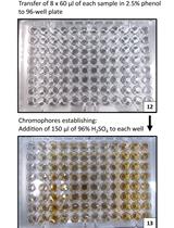
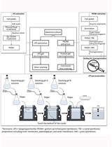
The presence of intracellular glycogen can be detected by the following iodine staining technique. Cells with glycogen stain dark brown, whereas in its absence they remain with a pale yellowish color. It is hypothesized that iodine atoms fit into helical coils formed by the α-polyglucan to form a coloured glycogen-iodine complex. Here, we have studied the expression of Streptococcus mutans (S. mutans) genes that control the biosynthesis of this polysaccharide (Asencion Diez et al., 2013). Thus, we expressed glgC and glgD genes coding for both ADP-Glc pyrophosphorylase subunits in Escherichia coli (E. coli) AC70R1-504 cells to complement the deficient accumulation of glycogen by this strain (Iglesias et al., 1993). In control cells or in those where an inactive protein was expressed, the synthesis of the polysaccharide was undetectable by this iodine staining technique.
Keywords: Reserve polysaccharide (储备多糖)Materials and Reagents
- Cells: Non-transformed E. coli AC70R1-504 or harboring plasmids with the S. mutans glgC and glgD genes, separately or combined
Note: This strain has a deficient production of the ADP-glucose pyrophosphorylase enzyme in absence of complementary plasmids (Morán-Zorzano et al., 2007).
- Luria-Bertani (LB) liquid medium
- Antibiotics: Kanamycin (US Biological, catalog number: K0010 ) and spectinomycin (Sigma-Aldrich, catalog number: S4014 )
- Inducers: Isopropyl β-D-1-thiogalactopyranoside (IPTG) (Sigma-Aldrich, catalog number: I6758 ) and nalidixic acid (Sigma-Aldrich, catalog number: N4382 )
- Plasmids: pMAB6/glgC, expressing S. mutans glgC (induced by nalidixic acid) and pMAB5/glgD, expressing S. mutans glgD (induced by IPTG)
Note: These plasmids are compatible and bear resistance to spectinomycin and kanamycin, respectively (Asencion Diez et al., 2013).
- D(+) Glucose (Sigma-Aldrich, catalog number: G5767 )
- Iodine crystals (Biopack Medical, catalog number: 2000162300 )
Equipment
- 1.5 ml microcentrifuge tube (Deltalab, catalog number: 200400 )
- Microcentrifuge (Beckman Coulter, model: 22R )
- Shaker (at least 200 rpm) at 37 °C (Thermo Fisher Scientific)
Procedure
文章信息
版权信息
© 2014 The Authors; exclusive licensee Bio-protocol LLC.
如何引用
Demonte, A. M., Diez, M. D. A., Guerrero, S. A., Ballicora, M. A. and Iglesias, A. A. (2014). Iodine Staining of Escherichia coli Expressing Genes Involved in the Synthesis of Bacterial Glycogen. Bio-protocol 4(17): e1224. DOI: 10.21769/BioProtoc.1224.
分类
微生物学 > 微生物生物化学 > 糖类
生物化学 > 糖类 > 糖原
细胞生物学 > 细胞染色 > 糖类
您对这篇实验方法有问题吗?
在此处发布您的问题,我们将邀请本文作者来回答。同时,我们会将您的问题发布到Bio-protocol Exchange,以便寻求社区成员的帮助。