

Mesenchymal Stem Cell (MSC) Aggregate Formation in vivo
间充质干细胞(MSC)聚合体的体内形成试验


发布: 2014年07月20日第4卷第14期 DOI: 10.21769/BioProtoc.1181 浏览次数: 12549
评审: Anonymous reviewer(s)
参见作者原研究论文
The authors used this protocol in:

Nov 2013


Abstract
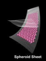
Human mesenchymal stem/progenitor cells (MSCs) isolated from various adult tissues show remarkable therapeutic potential and are being employed in clinical trials for the treatment of numerous diseases (Prockop et al., 2010). While routes of cell administration vary, profound beneficial effects of MSCs in animal models have been observed following intraperitoneal injections of the cells (Roddy et al., 2011). Similar to MSC spheres formed in culture under conditions where attachment to plastic is not permitted (Bartosh et al., 2010), MSCs injected into the peritoneum of mice spontaneously aggregate into 3D sphere-like structures (Bartosh et al., 2013). During the process of sphere assembly and compaction, MSCs upregulate expression of numerous therapeutic anti-inflammatory and immune modulatory factors. Here we describe the method we previously used for the generation of human bone marrow-derived MSC aggregates/spheres in vivo (Bartosh et al., 2013). By tagging the MSCs with green fluorescent protein (GFP), the aggregates formed can be easily visualized, collected and analyzed for changes in cellular properties and interactions with host immune cells.
Keywords: MSCs (骨髓间充质干细胞)Materials and Reagents
- Human bone marrow mesenchymal stem cells expressing green fluorescent protein (GFP-MSCs) from The Center for the Preparation and Distribution of Adult Stem Cells (http://medicine.tamhsc.edu/irm/msc-distribution.html)
- C57BL/6J or BALB/C mice (2-3 months of age) (The Jackson Laboratory)
- Phosphate-buffered saline (PBS) without Ca2+ and Mg2+ (pH 7.4) (Life Technologies, catalog number: 10010 )
- Hank’s Balanced Salt Solution (HBSS) without Ca2+ and Mg2+ (Lonza, catalog number: 04-315Q )
- 0.25% trypsin with 1x EDTA (Life Technologies, catalog number: 25200 )
- Minimum Essential Medium alpha (Life Technologies, catalog number: 12561 )
- Premium select fetal bovine serum (Atlanta Biologicals, catalog number: S11550 )
- Penicillin-streptomycin (Life Technologies, catalog number: 15140 )
- 100x L-glutamine (Life Technologies, catalog number: 25030 )
- Complete culture medium (CCM) for MSC growth (see Recipes)
Equipment
- Stericup-GP 0.22 µm vacuum filtration device (EMD Millipore, catalog number: SCGPU05RE )
- Water bath set to 37 °C
- Centrifuge with swinging-bucket rotor and adaptors for 50 ml conical tubes
- 50 ml sterile conical tube (BD Biosciences, Falcon®, catalog number: 352070 )
- Humidified cell culture incubator set to 37 °C and 5% CO2
- Upright microscope with 4x and 10x objectives and a filter set to visualize GFP
- 29 gauge needle with 1 ml syringe (Terumo Europe N.V., catalog number: 05M2913 )
- Isoflurane anesthesia system with nose cone for mouse
- Sterile dissecting scissors, pins, and curved forceps with a serrated edge
- Rubber or styrofoam platform
- Dissecting microscope with optional camera and monitor (Figure 1)
- Illumatool Bright Lights Systems LT 9900 with epi-fluorescence attachment (Lightools Research) and GFP filter set (Figure 1)
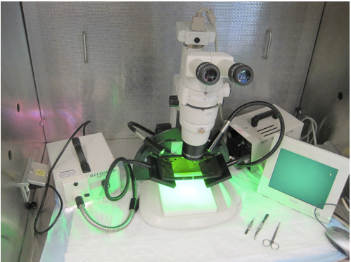
Figure 1. Equipment required to visualize and collect GFP-MSC aggregates/spheres from the mouse peritoneum. GFP-MSC aggregates/spheres can be visualized in the mouse peritoneum using a dissecting microscope with an epi-fluorescence attachment and GFP filter set. High quality images can be acquired with an appropriate camera mounted to the dissecting scope (a camera is not a requirement for collecting the aggregates).
Procedure
文章信息
版权信息
© 2014 The Authors; exclusive licensee Bio-protocol LLC.
如何引用
Bartosh, T. J. and Ylostalo, J. H. (2014). Mesenchymal Stem Cell (MSC) Aggregate Formation in vivo. Bio-protocol 4(14): e1181. DOI: 10.21769/BioProtoc.1181.
分类
干细胞 > 成体干细胞 > 间充质干细胞
干细胞 > 成体干细胞 > 维持和分化
细胞生物学 > 细胞分离和培养 > 3D细胞培养
您对这篇实验方法有问题吗?
在此处发布您的问题,我们将邀请本文作者来回答。同时,我们会将您的问题发布到Bio-protocol Exchange,以便寻求社区成员的帮助。