

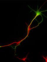
Neuron Culture from Mouse Superior Cervical Ganglion
小鼠颈上神经节中神经元的培养
发布: 2014年01月20日第4卷第2期 DOI: 10.21769/BioProtoc.1035 浏览次数: 15544
评审: Xuecai Ge
参见作者原研究论文
The authors used this protocol in:
Mar 2013


Abstract
The rodent superior cervical ganglion (SCG) is a useful and readily accessible source of neurons for studying the mechanisms of sympathetic nervous system (SNS) development and growth in vitro. The sympathetic nervous system (SNS) of early postnatal animals undergoes a great deal of remodeling and development; thus, neurons taken from mice at this age are primed to re-grow and establish synaptic connections after in situ removal. The stereotypic location and size of the SCG make it ideal for rapid isolation and dissociation. The protocol described here details the requirements for the dissection, culture and differentiation of SCG neurons. The protocol is suitable for culturing neurons from late embryonic gestation to approximately postnatal day 3. The culture technique discussed below utilizes glass coverslips for the microscopic examination of fixed cells.
Materials and Reagents
- Female mouse at desired gestational stage or early postnatal pups
- Sterile PBS (Corning Cellgro®, catalog number: 21-040-CV )
- L15 Leibovitz media (Corning Cellgro®, catalog number: 10-045-CV )
- Collagenase, Type 4 (1 mg) (Worthington Biochemical, catalog number: LS004182 )
- Collagenase enzyme solution (10 mg/ml in L15, filter-sterilized)
- Trypsin, 0.25% EDTA, Mg2+ Ca2+-free (Corning Cellgro®, catalog number: 25-053-Cl )
- Dulbecco’s Modified Eagle Medium (DMEM) (Corning Cellgro®, catalog number: 10-013-CV )
- Fetal bovine serum (FBS) (Equitech-Bio)
- Poly-D-lysine(PDL) (50 mg) (Sigma-Aldrich, catalog number: P0296 )
- Boric acid (Sigma-Aldrich, catalog number: B7660 )
- Sodium Tetraborate (Sigma-Aldrich, catalog number: B9876 )
- Laminin (1 mg) (BD Biosciences, catalog number: 354232 )
- 2.5 s Nerve Growth Factor (100 μg) (BD Biosciences, catalog number: 356004 )
- Concentrated nitric acid (Fisher Scientific, catalog number: A200-212 )
- Penicillin/streptomycin mix (Life Technologies, catalog number: 15140-122 )
- Sterile, deionized water
- Cytosine arabinoside (AraC) (Sigma-Aldrich, catalog number: C6645 )
- 0.1 M borate buffer (pH 8.5) (see Recipes)
- Regular plating medium (see Recipes)
Equipment
- Tissue culture incubator
- 35 mm or 6-well TC plates
- 35 mm tissue culture treated Petri dishes
- 100 mm Petri dishes
- 150 mm plastic Petri dish
- German glass coverslips, 25 mm (Electron Microscopy Sciences, catalog number: 72196-25 )
- Ceramic racks (Thomas Scientific, catalog number: 8542E40 )
- Basic gravity convection oven (VWR International, catalog number: 414005-108 )
- Silicon rubber dissection plates
- Scissors
- Fine tipped forceps
- 26 gauge needles
- Fire-polished, cotton-plugged, siliconized Pasteur pipets or Barrier tip 200
- Reduced bore siliconized Pasteur pipets
- Serological pipet
- Stereoscopic microscope
- Fume hood
- Water bath
- 37 °C incubator
Procedure
文章信息
版权信息
© 2014 The Authors; exclusive licensee Bio-protocol LLC.
如何引用
Readers should cite both the Bio-protocol article and the original research article where this protocol was used:
- Jackson, M. and Tourtellotte, W. (2014). Neuron Culture from Mouse Superior Cervical Ganglion. Bio-protocol 4(2): e1035. DOI: 10.21769/BioProtoc.1035.
-
Quach, D. H., Oliveira-Fernandes, M., Gruner, K. A. and Tourtellotte, W. G. (2013). A sympathetic neuron autonomous role for Egr3-mediated gene regulation in dendrite morphogenesis and target tissue innervation. J Neurosci 33(10): 4570-4583.
分类
神经科学 > 发育 > 神经元
神经科学 > 细胞机理 > 细胞分离和培养
您对这篇实验方法有问题吗?
在此处发布您的问题,我们将邀请本文作者来回答。同时,我们会将您的问题发布到Bio-protocol Exchange,以便寻求社区成员的帮助。