往期刊物2014

卷册: 4, 期号: 11

left

Dec

20

Dec

5

Nov

20

Nov

5

Oct

20

Oct

5

Sep

20

Sep

5

Aug

20

Aug

5

Jul

20

Jul

5

Jun

20

Jun

5

May

20

May

5

Apr

20

Apr

5

Mar

20

Mar

5

Feb

20

Feb

5

Jan

20

Jan

5

right

生物化学

Triton X-114法小规模提取疏水性蛋白

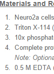
Small-scale Triton X-114 Extraction of Hydrophobic Proteins

Triton X-114法小规模提取疏水性蛋白

YT Yuzuru Taguchi
Hermann M.  Schätzl Hermann M. Schätzl
17947 Views
Jun 5, 2014
Here we introduce a protocol for Triton X-114 extraction which we used in our recently-published paper (Taguchi et al., 2013). It is a versatile method to concentrate or partially purify hydrophobic proteins. The presented protocol is based on the protocol published by Bordier (Bordier, 1981) but more simplified and down-scaled for more small-scale and simpler use (Taguchi et al., 2013).Triton X-114 (TX114) is a non-ionic detergent which has a relatively low clouding point at 22 °C and separates into detergent (Det) and aqueous (Aq) phase at temperatures above the clouding point. During phase separation, hydrophobic solutes in the TX114 solution are sequestered to the Det phase, while hydrophilic solutes are sequestered to the Aq phase. Utilizing this phenomenon, TX114 extraction is a very versatile technique to efficiently concentrate hydrophobic proteins, especially glycosylphosphatidylinositol (GPI)-anchored proteins like the prion protein (PrP), because they have substantial amounts of highly hydrophobic moieties. Besides, phase separation using TX114 tolerates a variety of conditions, e.g. different pH or relatively low concentrations of guanidine hydrochloride. Since the hydrophobic proteins are sequestered to the Det phase as long as the phase separation occurs, and if the hydrophobicity of the protein of interest is not affected by pH or denaturant, this technique can be also utilized to change buffers or to remove denaturants. When using enzymes or proteases which maintain activities in detergent solutions, TX114 can also be used to separate hydrophobic from the water-soluble hydrophilic moieties upon enzymatic digestion of proteins, as done by us using in vitro digestion of PrP with phosphatidylinositol-specific phospholipase C (Taguchi et al., 2013).
小鼠肺部细胞核蛋白制备法

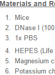
Protocol for Preparation of Nuclear Protein from Mouse Lungs

小鼠肺部细胞核蛋白制备法

ZX Zuoxiang Xiao
QJ Qun Jiang
14862 Views
Jun 5, 2014
This protocol describes how to extract nuclear protein from mouse lungs, including tissue preparation, stepwise lysis of cells and centrifugal isolation of nuclear protein fraction. This is an efficient method to get comparable nuclear protein extracts from lung tissues.
荧光热变分析测定假激酶配体的相互作用

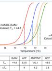
Determination of Pseudokinase-ligand Interaction by a Fluorescence-based Thermal Shift Assay

荧光热变分析测定假激酶配体的相互作用

IL Isabelle S Lucet
JH Joanne M. Hildebrand
PC Peter E. Czabotar
Jian-Guo Zhang Jian-Guo Zhang
NN Nicos A. Nicola
JS John Silke
JB Jeffrey J. Babon
JM James M. Murphy
12964 Views
Jun 5, 2014
This protocol describes a robust technique for the measurement of pseudokinase-ligand interaction by a fluorescence-based Thermal Shift Assay (TSA). Pseudokinases are kinase-like proteins that have recently emerged as crucial regulators of signal transduction and may therefore represent a novel class of drug targets. Unlike kinases, the catalytic efficiency of pseudokinases is rather poor or non existent, making it difficult to dissect the function of their nucleotide binding sites. Thermal denaturation-based methods have proven to be a powerful method for determining ligand binding capacity to purified pseudokinases and can inform on the potential drugability of the nucleotide binding site. This assay takes advantage of a change in flurorescence arising when a flurorescence dye, in this instance SYPRO® Orange, binds to hydrohobic patches that become exposed when a protein undergoes thermal denaturation. Ligand binding to a protein is known to increase its thermal stability which is reflected by a shift observed in the thermal denaturation curve between the unliganded protein and the liganded protein. This generalized protocol can also be tailored to other protein families. In addition, thermal denaturation-based methods can be used to identify optimal buffer conditions that may increase protein stability.

细胞生物学

蔗糖不连续密度梯度离心法进行小规模亚细胞分级分离

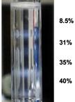
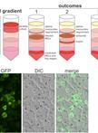
Small-scale Subcellular Fractionation with Sucrose Step Gradient

蔗糖不连续密度梯度离心法进行小规模亚细胞分级分离

YT Yuzuru Taguchi
Hermann M.  Schätzl Hermann M. Schätzl
16967 Views
Jun 5, 2014
Here, we introduce the protocol for small-scale and simple subcellular fractionation used in our recent publication (Taguchi et al., 2013), which uses homogenization by passing through needles and sucrose step-gradient. Subcellular fractionation is a very useful technique but usually a large number of cells are required. Because we needed subcellular fractionation of transiently-transfected cells, we developed a protocol for smaller numbers of cells. Our protocol for the subcellular fractionation is based on the protocol published by de Araújo and Huber (de Araujo et al., 2007), although substantial modifications have been made according to our experiences and information from personal communications. As optimal conditions seem to vary between cell lines, we advise to further modify the protocol to optimize for individual experiments. Our method is simple but sufficient for analysis of integral membrane proteins or proteins anchored to organelles by glycosylphosphatidylinositol or other lipid anchors, e.g. prion protein. However, proteins non-covalently attached to membranes or membrane proteins of organelles seem to be more prone to dissociation from the organelles during preparation and, if these proteins are the object of study, further modifications might be necessary. Unlike in a continuous gradient, where a protein of interest is scattered over a wide range, step-gradient fractionation is advantageous in detection of relatively small amounts of proteins from small-scale experiments, because it concentrates the protein of interest in one fraction, if an appropriate combination of sucrose concentrations is used.
研究异染色体和常染色质修复蛋白补充的特定位点诱导DNA氧化损伤新方法

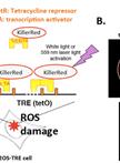
Novel Method for Site-specific Induction of Oxidative DNA Damage to Study Recruitment of Repair Proteins to Heterochromatin and Euchromatin

研究异染色体和常染色质修复蛋白补充的特定位点诱导DNA氧化损伤新方法

Leizhen  Wei Leizhen Wei
Satoshi  Nakajima Satoshi Nakajima
AL Arthur S. Levine
Li  Lan Li Lan
9689 Views
Jun 5, 2014
ROS-induced DNA damage is repaired in living cells within a temporal and spatial context, and chromatin structure is critical to a consideration of DNA repair processes in situ. It’s well known that chromatin remodeling factors participate in many DNA damage repair pathways, indicating the importance of chromatin remodeling in facilitating DNA damage repair. To date, there has been no method to induce site-specific oxidative DNA damage in living cells. Therefore, it is not known whether the DNA repair mechanisms differ within active or condensed chromatin. We recently established a novel method, DTG (Damage Targeted at one Genome-site), to study DNA damage response of reactive oxygen species (ROS)-induced DNA damage in living cell at one genome loci with active or inactive transcription. For this, we integrated a tetracycline responsive elements (TRE) cassette (~90 kb) at X-chromosome in U2OS cells (Lan et al., 2010), then fused KillerRed (KR), a light-stimulated ROS-inducer which can specifically produce ROS-induced DNA damage, to a tet-repressor (tetR-KR, OFF) or a transcription activator (TA-KR, ON) (Lan et al., 2014) (Figure 1). TetR-KR or TA-KR binds to the TRE cassette and induces ROS damage under hetero- or euchromatin states, respectively. How chromatin states regulate the DNA damage response processes can be examined by using this powerful method.

免疫学

清除小鼠巨噬细胞的方法

Protocol for Macrophage Depletion from Mice

清除小鼠巨噬细胞的方法

ZX Zuoxiang Xiao
QJ Qun Jiang
19264 Views
Jun 5, 2014
Macrophage depletion has been used extensively to study autoimmune disease and more recently in tumor models. The clodronate-containing liposomes will be recognized as foreign particles and get engulfed by macrophages upon dosing into the animal by the chosen routes. Consequently, macrophages that have engulfed liposomes will all be destroyed by the liposomal. In the protocol presented here the clodronate-containing liposomes were used to systemically deplete macrophages in mice.
体外炎性体试验

In vitro Inflammasome Assay

体外炎性体试验

Mario M.  Zaiss Mario M. Zaiss
KM Kendle M. Maslowski
14099 Views
Jun 5, 2014
Innate immune cells sense pathogen and danger-associated molecular patterns (PAMPs and DAMPs) through a range of innate immune pattern recognition receptors (PRRs). One type of PRRs are the Nod-like receptors (NLRs), which form inflammasomes; a molecular platform required for the recruitment and activation of Caspase-1, which in turn cleaves and activates IL-1β, IL-18. Examples of inflammasome forming NLRs are NLRP3, NLRP1, NAIP and NLRC4. We can easily identify new inflammasome activators by performing the following protocol.
鉴定蠕虫诱导产生的2型 CD4+T细胞和第2组先天淋巴细胞(ILC2)的方法

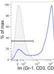
Identification of Helminth-induced Type 2 CD4+ T Cells and ILC2s

鉴定蠕虫诱导产生的2型 CD4+T细胞和第2组先天淋巴细胞(ILC2)的方法

Mario M.  Zaiss Mario M. Zaiss
KM Kendle M. Maslowski
8725 Views
Jun 5, 2014
After activation, T cells differentiate into different T helper (Th) subsets, namely Th1, Th2, and Th17. These different Th subsets are associated with the production of particular cytokines endowing them with different functions. In immunity against helminth infections the Th2 cell subset plays an important role. Th2 cells typically produce IL-4, IL-5, IL-13, and IL-9 resulting in antibody-isotype switching to IgE, eosinophilia, basophilia, mucin production, and smooth muscle cell hyperactivity. Here we analyze the development of the pathogen specific Th2 immune responses in mice after infection with the helminth parasite, Heligmosomoides polygyrus bakeri, the induction of innate lymphoid cells type 2 (ILC2) and the activation of the inflammasome in macrophages by excretory/secretory products of Heligmosomoides polygyrus bakeri.

微生物学

寄生虫蛋白质提取物的制备和蛋白印迹分析

Preparation of Parasite Protein Extracts and Western Blot Analysis

寄生虫蛋白质提取物的制备和蛋白印迹分析

AH Arlett Heiber
TS Tobias Spielmann
25557 Views
Jun 5, 2014
In order to prepare protein extracts of Plasmodium falciparum blood stages for western blot analysis, infected red blood cells (iRBC) need to be separated from uninfected red blood cells (uRBC) which make up the bulk of the parasite culture. Depending on the localisation of the parasite protein of interest, different methods are available to achieve this. If the protein is present within the parasite or is attached to a cellular structure of the iRBC cell, saponin can be used. This reagent lyses the membranes of infected and uninfected erythrocytes, the Maurer´s clefts (vesicular structures in the iRBC) and the parasitophorous vacuole membrane containing the parasite but leaves the parasite plasma membrane intact, providing a convenient procedure to isolate intact parasites without uRBCs. However, this method has the disadvantage that the host cell cytosol and the parasitophorous vacuole (PV) content of iRBCs are lost. If this has to be avoided, it is possible to use a Percoll gradient to separate intact iRBCs from uRBCs. Sequential treatment with Tetanolysin and saponin can then be used to selectively release the iRBC cytosol and the PV content from the parasite. These selective lysis methods are also suitable to determine the subcellular localisation of a protein of interest.
胞内糖原分析

Intracellular Glycogen Assays

胞内糖原分析

Yong  Jun  Goh Yong Jun Goh
TK Todd R. Klaenhammer
13074 Views
Jun 5, 2014
Glycogen, a soluble multi-branched glucose homopolysaccharide, is composed of chains of α-1,4-linked glucose residues interconnected by α-1,6-linked branches. The classical biosynthetic pathway involves phosphoglucomutase (Pgm), glucose-1-phosphate adenylyltransferase (GlgC or GlgCD), glycogen synthase (GlgA) and branching enzyme (GlgB). Phosphoglucomutase converts glucose-6-phosphate into glucose-1-phosphate, which serves as a substrate for ADP-glucose synthesis catalyzed by GlgC or GlgCD. Then, GlgA catalyzes the transfer of glucosyl units from ADP-glucose to the elongating chain of linear α-1,4-glucan. GlgB subsequently cleaves off portions of the glucan and links it to internal glucose molecules in existing chains via α-1,6 glycosidic bonds to form the glycogen structure. Glycogen breakdown is mediated by glycogen phosphorylase (GlgP) and debranching enzyme (GlgX), which catalyze the sequential phosphorolysis of α-1,4-glucosyl linkages in the glucan chain from the non-reducing ends and debranching of the limit dextrins generated by GlgP, respectively. An increasing number of studies have revealed the involvement of glycogen metabolism in a multitude of physiological functions in some prokaryotes beyond the function of synthesizing energy storage compounds. Lactobacillus acidophilus NCFM was the first probiotic lactic acid bacterium demonstrated to possess a functional glycogen biosynthesis pathway that is involved in its growth, bile tolerance and complex carbohydrate metabolism (Goh and Klaenhammer, 2013). The following qualitative (for detection of intracellular glycogen) and quantitative (for measurement of intracellular glycogen content) intracellular glycogen assay protocols for Lactobacillus acidophilus (L. acidophilus) were modified from previous works (Govons et al., 1969; Law et al., 1995; Parrou and Francois, 1997) and should be applicable to other lactic acid bacteria as well as most microorganisms.
幼兔肠道中不同菌株的定植竞争分析

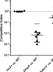
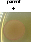
Infant Rabbit Colonization Competition Assays

幼兔肠道中不同菌株的定植竞争分析

Yang  Fu Yang Fu
JM John J. Mekalanos
10435 Views
Jun 5, 2014
Enteric pathogenic bacteria such as Vibrio cholerae and enteropathogenic Escherichia coli (E. coli) cause life-threatening diarrheal diseases that have afflicted humans for centuries. Understanding the effectors required for intestinal colonization is very important to research on bacteria pathogenesis, and is also important to testing new therapeutics and development of the novel vaccines. Here, we describe the Infant Rabbit Colonization Competition Assay, a variant method of the powerful, nonsurgical animal model reported by Ritchie et al. (2010). In our modified assay, wild type and mutant strains are mixed together and inoculated into 2-day-old New Zealand white rabbits. The competitive index for each mutant measures the colonization capacity of the mutant relative to its wild type parental strain in the gastrointestinal tract. Compared to the traditional Sucking Mice model, the clinical and histologic signs of Vibrio cholerae (V. cholerae)-induced disease of infant rabbits more closely resemble human cholera. The larger input bacteria amount of this model also facilitates high-throughput screens, such as Tn-Seq technology (Fu et al., 2013).
PF13_0191-GFP 表达寄生虫的免疫透射电子显微分析

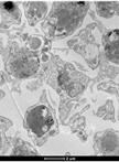
Immuno-EM Analysis of PF13_0191-GFP Expressing Parasites

PF13_0191-GFP 表达寄生虫的免疫透射电子显微分析

AH Arlett Heiber
SR Silke Retzlaff
TS Tobias Spielmann
9608 Views
Jun 5, 2014
This protocol was used to prepare pre-embedding samples of Plasmodium falciparum blood stage parasites that overexpressed the parasite protein PF13_0191 tagged with GFP. Using GFP-specific antibodies and Protein A-Gold the localisation of the overexpressed protein in the infected host cell was determined using standard transmission electron microscopy (EM). Pre-embedding EM is a common method where the antibodies are introduced before embedding and sectioning. This method avoids the problem that antigens are often difficult to detect on EM-sections after embedding. In the method described here antigens in the parasite-infected host cell are detected. Entry of the antibody is made possible through permeabilisation of the host cell with tetanolysin. In principle this method could also be used to detect antigens within the parasite if the sample is appropriately fixed and permeabilised before addition of the relevant antibody. While access of the antibody will avoid the detection problems often seen with post-embedding methods, this procedure will produce comparably poorer morphology.
海豚葡萄球菌的QS抑制化合物的纯化和结构分析

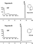
Purification and Structural Analysis of QS-inhibiting Compounds from Staphylococcus delphini

海豚葡萄球菌的QS抑制化合物的纯化和结构分析

YC Ya-Yun Chu
MN Mulugeta Nega
FG Friedrich Götz
8068 Views
Jun 5, 2014
The knowledge that many pathogens rely on cell-to-cell communication mechanisms known as quorum sensing, opens a new disease control strategy: quorum quenching. Here we present a purification protocol for molecules excreted by a group of Gram-positive zoonotic pathogen bacteria, the ‘Staphylococcus intermedius group’, that suppress the quorum sensing signaling and inhibit the growth of a broad spectrum of Gram-negative beta- and gamma-proteobacteria. These compounds were isolated from Staphylococcus delphini (S. delphini). They represent a new class of quorum quenchers with the chemical formula N-[2-(1H-indol-3-yl)ethyl]-urea and N-(2-phenethyl)-urea, which we named yayurea A and B, respectively. These substances can be isolated and purified from the culture supernatant using this upscalable purification method.

干细胞

竞争性骨髓移植

Competitive Bone-marrow Transplantations

竞争性骨髓移植

MM Maria Maryanovich
AG Atan Gross
25640 Views
Jun 5, 2014
Competitive bone marrow transplantation assay measures multi-lineage reconstitution of hematopoiesis in irradiated transplant recipient mice. Thus this assay is routinely used to determine haematopoietic stem and progenitor cells (HSPCs) functionality in vivo. The principle of the method is to transplant bone marrow donor cells (derived from transgenic mice of choice) on C57BL6 background together with normal competitor bone marrow. In order to distinguish donor from competitor cells upon transplantation, usually competitor mice are congenic and carry the differential B cell antigen originally designated Ly5.1 and CD45.1.A typical competitive bone marrow transplantation experiment will contain two transplantation groups, donor (transgenic mice of choice and their controls) are transplanted in competition with normal competitors and engraftment efficiency is evaluated in both blood and bone marrow.