Isolation and Culture of Peritoneal Cell-derived Mast Cells
来源于腹水的肥大细胞分离和培养
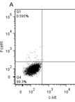
The generation of mast cells for in vitro studies comes from a variety of sources including mast cell lines (MC/9) (McCurdy et al., 2001), bone marrow-derived mast cells (BMMCs) (Supajatura et al., 2001), skin-derived mast cells (FSMCs) (Matsushima et al., 2004), peritoneal-derived mast cells (PMCs) (Hochdorfer et al., 2011) and peritoneal cell-derived cultured mast cells (PCMCs) (Vukman et al., 2012). PCMCs are generally used for in vitro studies because they are a more mature source of mast cells when compared to mast cells generated or obtained from other sources. They can differ, for example, in their pro-inflammatory responses to bacterial antigens and toll like receptors (TLRs) ligands (Mrabet-Dahbi et al., 2009). In comparison to BMMCs [see the protocol “Isolation and Culture of Bone Marrow-derived Mast Cells” (Vukman et al., 2014)] or mast cell lines they express a wider range of TLRs, and secrete significantly more cytokines when stimulated with TLR ligands (Mrabet-Dahbi et al., 2009). Therefore, when examining pro-inflammatory responses, mast cells generated from cells obtained from the peritoneal cavity give stronger responses. PCMCs can also be generated from knockout and transgenic mice making them a good source to examine specific factors important for mast cell function. However, due to the low yield of cells generated using this method (1 million per mouse) their use is restricted and therefore in most studies more than one source of mast cells may be required. The different sources of mast cells can display phenotypical and functional differences and therefore it is important that when designing an experiment, the correct cellular source is obtained. Here, we describe a protocol for the isolation and culture of murine mast cells from peritoneal cavity cells.