往期刊物2013

卷册: 3, 期号: 22

left

Dec

20

Dec

5

Nov

20

Nov

5

Oct

20

Oct

5

Sep

20

Sep

5

Aug

20

Aug

5

Jul

20

Jul

5

Jun

20

Jun

5

May

20

May

5

Apr

20

Apr

5

Mar

20

Mar

5

Feb

20

Feb

5

Jan

20

Jan

5

right

细胞生物学

小鼠腹水中巨噬细胞的采集和培养

Harvest and Culture of Mouse Peritoneal Macrophages

小鼠腹水中巨噬细胞的采集和培养

Mingfang  Lu Mingfang Lu
AV Alan W. Varley
38982 Views
Nov 20, 2013
Peritoneal macrophages are used as primary macrophages in lots of studies, mainly because they are easy to obtain. Injection of thioglycollate broth i.p. induces inflammatory responses and elicits large numbers of macrophages. This protocol can be used for harvesting resident or thioglycollate-elicited peritoneal cells. Peritoneal macrophages are non-adherent in situ and when they are cultured in dishes, they become adherent so that macrophages may be separated from other types of cells in peritoneal cavity.

免疫学

胃内容物的气管吸入模型

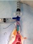
Gastric Aspiration Models

胃内容物的气管吸入模型

Bruce  A.  Davidson Bruce A. Davidson
RA Ravi Alluri
19735 Views
Nov 20, 2013
The procedures described below are for producing gastric aspiration pneumonitis in mice with alterations for rats and rabbits described parenthetically. We use 4 different injury vehicles delivered intratracheally to investigate the inflammatory responses to gastric aspiration:1) Normal saline (NS) as the injury vehicle control2) NS + HCl, pH = 1.25 (acid)3) NS + gastric particles, pH ≈ 5.3 (part.)4) NS + gastric particles + HCl, pH = 1.25 (acid + part.)The volume, pH, and gastric particle concentration all affect the resulting lung injury. In mice, we generally use an injury volume of 3.6 ml/kg (rat: 1.2 ml/kg, rabbit: 2.4 ml/kg), an injury pH (for the acid-containing vehicles) of 1.25, and a gastric particulate concentration (in the particulate-containing vehicles) of 10 mg/ml (rat: 40 mg/ml). In our hands this results in a maximal, non-lethal lung injury with ≤ 10% mortality for the most injurious vehicle (i.e., acid + part.) The maximum tolerable particulate concentration needs to be determined empirically for any new strains to be used, especially in genetically-altered mice, because an altered inflammatory response may have detrimental affects on mortality.We have extensive experience utilizing these procedures in the outbred strain, CD-1, as well as many genetically-altered inbred stains on the C57BL/6 background. Choice of strain should be carefully considered, especially in terms of strain-specific immune bias, to assure proper data interpretation. The size of the mouse should be ≥ 20 g at the time of injury. Smaller mice can be attempted, if necessary, but the surgical manipulation becomes increasingly more difficult and the surgery survival rate decreases substantially. There are no size or strain constraints for rat and rabbit models, but we generally use Long-Evans rats at 250-300 g and New Zealand White rats at ≈ 2 kg at the time of initial injury.
采用流式细胞术评估人树突细胞的抗原摄取

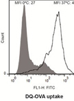
Assessment of Human Dendritic Cell Antigen Uptake by Flow Cytometry

采用流式细胞术评估人树突细胞的抗原摄取

AL Ana Luque
SC Sonia Cárdenas-Brito
RO Rut Olivar
JA Josep M. Aran
18037 Views
Nov 20, 2013
Antigen uptake by dendritic cells is the first key step towards induction of antigen-specific T-cell responses. This flow cytometry-based protocol describes the analysis of dendritic cell uptake of soluble antigens through two different mechanisms: non-specific macropinocytosis (using Lucifer Yelloy CH), and receptor-mediated endocytosis (using DQTM Ovalbumin). The protocol is generated based on data presented in Olivar et al. (2013).
免疫细胞化学法检测曼氏血吸虫孢囊中的重组毒素

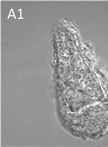
Immunocytochemical Detection of Recombinant Biomphalysin on Schistosoma mansoni Sporocysts

免疫细胞化学法检测曼氏血吸虫孢囊中的重组毒素

David  Duval David Duval
RG Richard Galinier
JP Julien Portela
GM Guillaume Mitta
BG Benjamin Gourbal
8819 Views
Nov 20, 2013
Schistosomiasis, or bilharzia, is a tropical disease caused by worms of the genus Schistosoma which infect about 200 million people. The life cycle of the parasite requires Biomphalaria, a specific genus of freshwater snails, as intermediate. Using an interactome approach employing B. glabrata plasma and S. mansoni primary sporocyst extracts, we identified a new cytolytic protein called Biomphalysin that displays similarities to members of the β-PFT superfamily known to form channels in targeted membranes. To investigate its mechanism of action, we produced a recombinant protein flanked by an N-terminal 6 histidine tag. Then, we investigated the ability of Biomphalysin to interact with the sporocyst tegument. This optimized protocol describes an immunocytochemical procedure to detect histidine tagged recombinant protein on the sporocyst tegumental membrane.

微生物学

小鼠血管通透性诱导的分析评估

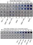
Assay to Evaluate Vascular Permeability Induction in Mice

小鼠血管通透性诱导的分析评估

HP Henry Puerta-Guardo
AR Arturo Raya-Sandino
LG Lorenza González-Mariscal
VR Victor H. Rosales
JA José Ayala-Dávila
BC Bibiana Chávez-Mungía
DM Daniel Martínez-Fong
FM Fernando Medina
Juan  E.  Ludert Juan E. Ludert
Rosa María del  Angel Rosa María del Angel
22341 Views
Nov 20, 2013
Dengue virus infection usually courses as a benign self-limited fever, called dengue fever. However, on occasions it can progress to a life-threatening complication known as severe dengue (SD). A hallmark of SD is a sharp increase in vascular permeability. Secondary infections are considered a risk factor to develop SD, presumably through a mechanism called Antibody-Dependent Enhancement (ADE) of infection in cells with the capacity to bind antigen-antibody complexes, such as macrophages, and to trigger a subsequent aberrant cytokine response. The massive release of cytokine from macrophages has been postulated to cause changes in vascular permeability. The vascular permeability assay presented in this protocol is designed to assess whether any compound or cell-secreted product or soluble factor present in sera from patients may induce plasma leakage in mice. This test was used in the laboratory to determine whether cytokines and soluble factors produced in vitro by macrophages infected with dengue virus or dengue virus in the presence of facilitating antibodies are able to induce plasma leakage in vivo. Macrophages were infected with dengue virus or dengue virus in the presence of facilitating antibodies for 48 h. After this time, the conditioned supernatant containing cytokines and soluble factors released by the macrophages were collected and inoculated intraperitoneally into CD-1 mice. Twenty four hours after the first inoculation, mice were reinoculated with a second dose with Evans blue dye. After another 24 h, mice were euthanized and the amount of Evans blue present in the blood and lung was determined by spectrophotometric analysis. The assay was able to show differences in the capacity of the conditioned media to induce vascular permeability changes in the inoculated animals (Puerta-Guardo et al., 2013).
体内和体外分离和检测一种百日咳杆菌反应调节蛋白BvgA的磷酸化和非磷酸化形式

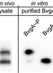
Separation and Detection of Phosphorylated and Nonphosphorylated BvgA, a Bordetella pertussis Response Regulator, in vivo and in vitro

体内和体外分离和检测一种百日咳杆菌反应调节蛋白BvgA的磷酸化和非磷酸化形式

QC Qing Chen
AB Alice Boulanger
DH Deborah M. Hinton
SS Scott Stibitz
11185 Views
Nov 20, 2013
Protein phosphorylation plays a central role in signal transduction in bacteria. However, separation and detection of the phosphorylated protein from its nonphosphorylated form remain challenging. Here we describe a method to detect phosphorylation of the Bordetella pertussis response regulator BvgA, which is phosphorylated at an aspartate residue (Boulanger et al., 2013). This method is based on the proprietary adduct, Phos-tagTM, a dinuclear metal complex, which together with Zn2+ or Mn2+, forms a complex with a phosphomonoesterdianion, such as the phosphorylated aspartate of a response regulator (Barbieri and Stock, 2008; Kinoshita and Kinoshita-Kikuta, 2011). For in vivo detection, B. pertussis cells are lysed in mild formic acid at 4 °C to minimize the disruption of the phospho-aspartate bond, and the phosphorylated BvgA is separated from its nonphosphorylated form by electrophoresis (SDS-PAGE) containing Phos-tagTM. Both forms of BvgA are subsequently detected by Western Blot analysis. Quantification of the level of phosphorylated BvgA formed after treatment with acetyl phosphate in vitro is also easily accomplished. Thus, this technique allows one to readily assess the levels of BvgA phosphorylation in B. pertussis and in E. coli under different laboratory conditions in vivo or after phosphorylation under varying reaction conditions in vitro (this research was supported in part by the Intramural Research Program of the NIH, NIDDK).
志贺氏杆菌IPaD和IPAB表面定位

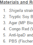
Shigella IpaD and IpaB Surface Localizations

志贺氏杆菌IPaD和IPAB表面定位

Lionel  Schiavolin Lionel Schiavolin
AM Alaeddine Meghraoui
AA Abdelmounnaim Allaoui
10582 Views
Nov 20, 2013
Shigella uses a type III secretion system to invade host cell and to cause disease. Secretion control and insertion of a translocation pore into cell membrane are critical steps for pathogenesis and are tightly linked to the formation of the needle tip complex formed by the IpaB and IpaD proteins (Veenendaal et al., 2007). Surface localizations of IpaD and IpaB were monitored by FACS analysis according to the localization protocol for Pseudomonas aeruginosa homolog PcrV (Lee et al., 2010).
巢状曲霉转运蛋白动力学分析实验

Transport Assays in Aspergillus nidulans

巢状曲霉转运蛋白动力学分析实验

Emilia  Krypotou Emilia Krypotou
George  Diallinas George Diallinas
8802 Views
Nov 20, 2013
Transport assays allow the direct kinetic analysis of a specific transporter by measuring apparent Km and Vmax values, and permit the characterization of substrate specificity profiles through competition assays. In this protocol, we describe a rapid and easy method for performing uptake assays in the model filamentous ascomycete Aspergillus nidulans. These assays make use of A. nidulans germinating conidiospores, thus avoiding technical difficulties associated with the use of mycelia. The ease of construction genetic null mutants in this model fungus permits the rigorous characterization of any transporter in the absence of similar transporters with overlapping specificities, a common problem in relevant studies.

神经科学

在脊椎动物脑部采用荧光标记新鲜细胞核进行细胞周期分析

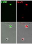
Cell Cycle Analysis in the Vertebrate Brain Using Immunolabeled Fresh Cell Nuclei

在脊椎动物脑部采用荧光标记新鲜细胞核进行细胞周期分析

Noelia  Lopez-Sanchez Noelia Lopez-Sanchez
Jose M.  Frade Jose M. Frade
17283 Views
Nov 20, 2013
Flow cytometry, a standard technique used for quantitative analysis of isolated cells, is routinely employed by immunologists and oncologists to study DNA content, protein expression, and other functional parameters in blood and tumor cells. Unfortunately, the use of this technique by neurobiologists has been hampered by the complexity of the nervous system, whose constituting cells can hardly be dissociated to obtain samples of sufficient quality. We have developed a simplified and quick method to purify and immunolabel cell nuclei with high sensitivity and low background. Our protocol allows the discrimination of single nuclei from doublets and larger aggregates, obtaining low coefficients of variation for cell cycle analysis with propidium iodide. In addition, due to the reduced sample handling this method has high recovery and good reproducibility. As an example, in this protocol we describe the isolation of cell nuclei from adult cerebral cortex, which are subsequently immunostained with antibodies against NeuN (a general neuronal marker) and EGR1 (an early response gene expressed by functionally active neurons), and subjected to flow cytometric gating and analysis. Nevertheless, the protocol can also be applied to other neural tissues from adult and embryonic brain.

植物科学

EMSA实验(凝胶迁移试验)分析RNA-蛋白质的相互作用

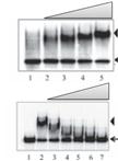
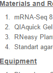
Analysis of RNA-protein Interactions Using Electrophoretic Mobility Shift Assay (Gel Shift Assay)

EMSA实验(凝胶迁移试验)分析RNA-蛋白质的相互作用

SP Saiprasad Goud Palusa
Anireddy  S. N.  Reddy Anireddy S. N. Reddy
30248 Views
Nov 20, 2013
RNA binding proteins (RBPs) play a crucial role in regulating gene expression at the post-transcriptional level at multiple steps including pre-mRNA splicing, polyadenylation, mRNA stability, mRNA localization and translation. RBPs regulate these processes primarily by binding to specific sequence elements in nascent or mature transcripts. There are several hundreds of RBPs in plants, but the targets of most of them are unknown. A variety of experimental methods have been developed to identify targets of an RBP. These include RNA immunoprecipitation (RIP), UV cross-linking and immunoprecipitation (CLIP) and many variations of CLIP (e.g. PAR-CLIP, iCLIP). These approaches depend on immunoprecipitation of RNAs bound to a specific RBP using an antibody to that RBP. Electrophoretic mobility shift assay (EMSA), also called gel shift assay, has been used to analyze protein-nucleic acid interactions. It is a simple and powerful method to analyze protein-RNA/DNA interactions. In RNA EMSA, RNA-protein complexes are visualized by comparing the migration of RNA in the presence of a protein. Generally, in RNA EMSA a specific RNA sequence is used to analyze its interaction with a protein. In vitro transcribed 32P labeled or chemically synthesized RNA with a fluorescent tag is incubated with or without the protein of interest and the reaction mixture is then run on native polyacrylamide gel electrophoresis. RNA-Protein complexes migrate slowly as compared to free RNA, which can be visualized using an imaging system. In addition to test binding of an RBP to RNA, EMSA is also used to map the region in RNA and/or protein that is involved in interaction. Furthermore, the binding affinity can also be quantified using EMSA.
水稻原生质体中的(双分子荧光互补)BiFC实验

Bimolecular Fluorescence Complementation (BIFC) Protocol for Rice Protoplast Transformation

水稻原生质体中的(双分子荧光互补)BiFC实验

KW Kun Wang
YL Ying Liu
Shaoqing  Li Shaoqing Li
16187 Views
Nov 20, 2013
After the plant cells are removed the cell walls by digestive enzyme, the plant protoplasts still have good cell activity. The protoplasts can be used to transiently express proteins of target genes in living plant cells through polyethylene glycol (PEG) mediated transformation. The purpose of this method is to employ the rice protoplasts and Green fluorescent protein (GFP) as an experimental system to observe the protein interactions in vivo. Meanwhile a 505~530 nm emission filter is used in confocal microscope to eliminate the interference of the autofluorescence from plant cells. The phenomenon of plant cell body spontaneous fluorescence can be eliminated by confocal observation.
黑杨的莽草酸羟基肉桂转移酶(HCT)活性分析

Shikimate Hydroxycinnamoyl Transferase (HCT) Activity Assays in Populus nigra

黑杨的莽草酸羟基肉桂转移酶(HCT)活性分析

Igor  Cesarino Igor Cesarino
RV Ruben Vanholme
GG Geert Goeminne
BV Bartel Vanholme
WB Wout Boerjan
14419 Views
Nov 20, 2013
Lignin is a complex phenolic polymer deposited in secondarily-thickened plant cell walls. The polymer is mainly derived from the three primary monolignols: p-coumaryl, coniferyl and sinapyl alcohol which give rise to p-hydroxyphenyl, guaiacyl and syringyl units (H, G and S units, respectively) when coupled into the polymer. The building blocks differ in their degree of methoxylation and their biosynthetic pathway is catalyzed by more than 10 enzymes. HCT plays a crucial role by channeling the phenylpropanoids towards the production of coniferyl and sinapyl alcohols. Interestingly, HCT has been reported to be implicated in the pathway both upstream and downstream of the 3-hydroxylation of the aromatic ring of p-coumaroyl shikimate (Figure 1) (Hoffmann et al., 2003; Hoffmann et al., 2004; Vanholme et al., 2013b). These features highlight the importance of developing an assay to reliably measure HCT activity in planta. Here, we describe a UPLC-MS-based method for the analysis of HCT activity in xylem total protein extracts of Populus nigra, which can be adapted to other woody and herbaceous plant species. The protocol was initially described in Vanholme et al. (2013a).
胞质蛋白与细胞骨架相互作用研究

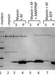
Binding Assay of Cytosolic Proteins to the Cytoskeleton

胞质蛋白与细胞骨架相互作用研究

Stefano   Del Duca Stefano Del Duca
Giampiero  Cai Giampiero Cai
12521 Views
Nov 20, 2013
Many cellular proteins interact with the cytoskeleton (both actin filaments and microtubules), either dynamically or permanently. This interaction is required during different aspects of the cell life, for example during the process of cell division. In addition, many enzymes interact transiently with actin filaments and microtubules in order to promote their cellular distribution. Several substances with inhibitory capacity can affect this binding and cause damages to cells. This protocol allows to analyze whether a protein interacts with either actin filaments or microtubules and, when applicable, the conditions controlling this interaction.The test is based on the specific binding between the protein of interest and the cytoskeletal filaments. As shown schematically in the diagram of example (see below), the test starts from the cell lysate to which actin filaments (produced from monomeric actin) are added. The mixture (performed under different experimental conditions chosen by the operator) is then incubated so that the protein of interest (in the example, myosin) binds to actin filaments. The sample is then centrifuged in order to separate unbound or weakly-bound proteins from actin filaments to which both the protein of interest and, eventually, traces of less specific proteins are associated.
早期结瘤时期的苜蓿中的转录组分析(Illumina测序)

Mapping and Analysis of Illumina Reads for Transcriptome of Medicago Truncatula During the Early Organogenesis of the Nodule

早期结瘤时期的苜蓿中的转录组分析(Illumina测序)

AB Alexandre Boscari
AF Alberto Ferrarini
JG Jennifer del Giudice
LV Luca Venturini
MD Massimo Delledone
AP Alain Puppo
10807 Views
Nov 20, 2013
Medicago truncatula serves as a model plant for legume genetics and genomics. We used RNA-Seq to characterize the transcriptome during the early organogenesis of the nodule and during its functioning. We generated approximately 135.5 million high-quality 36-bp reads, which were then aligned with the M. truncatula genome sequence (Mt3.0 version) and with sequences from a custom splice-junction database, for the detection of transcribed regions and splicing sites. Mapping and analysis of the reads conducted to the detection of 37,333 expressed transcription units (TUs), 1,670 had never been described before and were functionally annotated. We identified 7,595 new transcribed regions, mostly corresponding to 5’ and 3’ UTR extensions and new exons associated with 5,264 previously annotated genes. We also assessed the complexity in the nodulation transcriptome by performing a Cufflinks analysis to determine the frequency of the various alternatively spliced forms. Thus, we identified 23,164 different transcripts derived from 6,587 genes. Finally, we carried out a differential expression analysis, which provided a comprehensive view of transcriptional reprogramming during nodulation.All Illumina sequence data have been deposited in the NCBI short-read archive, and Sanger-sequenced PCR products have been deposited in GenBank (SRA048731). Assembled contigs longer than 200 bp have been deposited at TSA (JR366937-JR375780). Coverage data are available at http://ddlab.sci.univr.it/cgi-bin/gbrowse/medicago/.
棉花胚珠的培养和分析

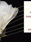
Cotton Ovules Culture and Analysis

棉花胚珠的培养和分析

Jiafu  Tan Jiafu Tan
FD Fenglin Deng
WT Wenxin Tang
JH Jie Han
GK Guo Kai
LT Lili Tu
XZ Xianlong Zhang
10641 Views
Nov 20, 2013
The cotton ovules culture was innovated by Beasley and Ting (1973), and named after them. It is a convenient system to analyze the effect of chemical or environmental treatment on fiber development directly on ovules. This protocol was generated according to previous published papers and our practical experience.