

往期刊物2012
卷册: 2, 期号: 6
细胞生物学
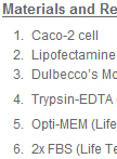
Stable Caco-2 Cell Line Construction
稳定Caco-2细胞系的建立

This protocol describes the construction of Caco-2 stable cell lines using the Lipofectamine transfection method.
免疫学
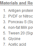
Antibody Purification from Western Blotting
利用免疫印迹法(WB)进行抗体纯化
This protocol describes a method of purifying antibodies from sera with denatured antigens immobilized on western blot membranes. Advantages include (1) fast and easy; (2) purification of antibody with antigen in denatured form allows high yield in case antigen protein solubility is limited. Disadvantage is that possible antibodies that recognize certain 3D structure in solution of antigens might not be purified using such a method. Regarding this issue, the flow through is recommended to be kept and can be used for other purification methods with folded antigens.
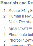
INF-gamma Release ELISpot Assay
酶联免疫斑点法(ELISpot)测定INF-γ释放水平
IFN-γ (Interferon-gamma) is produced mainly by activated T cells and NK cells. Production of IFN-γ (Interferon-gamma) by helper T cells as well as cytotoxic T cells is a hallmark of the TH1-type phenotype, thus, high-level production of IFN-γ (Interferon-gamma) is typically associated with effective host defense against intracellular pathogens. The Enzyme-Linked ImmunoSpot (ELISpot) assay is commonly used to assess the function of antigen specific T cells by detecting IFN-γ release. The ELISpot assay is a very sensitive immunoassay, allowing the detection of a secreted cytokine at the single cell level. With detection levels that can be as low as one cell in 100,000, the ELISpot is one of the most sensitive cellular assays available. Depending on the substance analyzed, the ELISpot assay is between 20 and 200 times more sensitive than a conventional ELISA.
分子生物学
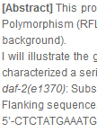
PCR-RFLP Genotyping of Point Mutations in Caenorhabditis elegans
线虫点突变的PCR-RFLP基因型鉴定

This protocol describes the basic principle of PCR/restriction digest genotyping of point mutations in worms, based on the principle of Restriction Fragment Length Polymorphism (RFLP) analysis. This type of genotyping is, particularly, useful when phenotypic analysis of animals carrying point mutations is difficult (e.g., in a complex genetic background). I will illustrate the general procedures, using an example of daf-2 gene, encoding the sole insulin/IGF-1 receptor of C. elegans. Gems et al.(1998) did a very elegant job and characterized a series of mutations of daf-2, including the following two temperature-sensitive hypomorphic alleles: daf-2(e1370): Substitution C/T (wild type/mutant), amino acid change: Missense P to SFlanking sequences: 5’-CTCTATGAAATGGTTACACTCGGTGCTCAGCATATATTGGTTTGAGTAATGAGGTGTIntracellular kinase domain, Class I, strong phenotype. daf-2(e1368): Substitution C/T (wild type/mutant), amino acid change: Missense S to LFlanking sequences: 5’-TCCGGAATTTACGTATTGAGGCAAAGTACTGTTCAGAAATVTATATGCTATCACAGTExtracellular ligand binding domain, Class II, weak phenotype. Here I will show you how to design the primers for PCR-RFLP analysis. daf-2(e1370): Designed by Seung-Jae Lee from the Kenyon labForward primer: 5’-CGGGATGAGACTGTCAAGATTGGAGATTTCGG-3’Reverse primer: 5’-CAACACCTCATCATTACTCAAACCAATCCATG-3’On the (-) strand, the nucleotide next to the 3’ end of reverse primer is G in wild-type allele, which is mutated to T in daf-2(e1370). Thus, by introducing another mutation (double C here, highlighted) into the reverse primer, it creates an Nco I-restriction site (i.e., CCATGG) only for PCR products derived from wild-type but NOT daf-2(1370). daf-2(e1368): Designed by Peichuan Zhang from the Kenyon labForward primer: 5’-GTTCCGGAATTTACGACGTATTGAGGCAACG-3’Reverse primer: 5’-CTATCGGATCGAGTGGTATATTTAAC-3’Similarly, on the (+) strand, the nucleotides next to the 3’ end of forward primer are TC in wild-type allele, and TT in daf-2(e1368). Thus, by introducing another mutation (C here, highlighted) into the forward primer, a restriction site of Acl I (i.e., AACGTT) is generated in the presence of daf-2(1368) point mutation. The key is to introduce new mutation(s) at the 3’ end of one of your primers. Since the difference of the sizes of digest products is just ~30-bp, the length of the primer, you have to pick the other primer to generate an amplicon of ~200-bp – 250-bp or so. Here is a website that can help you design the primers with appropriate restriction site for genotyping: http://helix.wustl.edu/dcaps/dcaps.html (dCAPS Finder 2.0) (Neff et al., 2002).