往期刊物2016

卷册: 6, 期号: 9

left

Dec

20

Dec

5

Nov

20

Nov

5

Oct

20

Oct

5

Sep

20

Sep

5

Aug

20

Aug

5

Jul

20

Jul

5

Jun

20

Jun

5

May

20

May

5

Apr

20

Apr

5

Mar

20

Mar

5

Feb

20

Feb

5

Jan

20

Jan

5

right

癌症生物学

用成纤维细胞对胃癌细胞进行三维凝胶侵袭试验

3D Gel Invasion Assay of Gastric Cancer Cells with Fibroblasts

用成纤维细胞对胃癌细胞进行三维凝胶侵袭试验

Masamitsu Tanaka Masamitsu Tanaka
12528 Views
May 5, 2016
Cancer tissue is composed of cancer cells and a large number of stromal cells including fibroblasts. In order to understand the relationship between fibroblasts and cancer cells during invasion of the stroma, 3D gel invasion assay is useful. Most tumors are associated with a biologically active type of fibroblasts known as cancer-associated fibroblasts (CAFs), which promote the invasion of cancer cells. Here, we describe the method of imaging the invasion by fluorescently labeled CAFs and gastric cancer cells in gels containing extracellular matrix. For two-color fluorescence labeling of living cells, long-chain dialkylcarbocyanines, DiO and DiI were used. This method is also applicable for studying invasion by other stromal cells and cancer cells, and for evaluation of drugs targeting cancer stromal cells.

发育生物学

果蝇幼虫和蛹中气管的制备和免疫荧光染色

Preparation and Immunofluorescence Staining of the Trachea in Drosophila Larvae and Pupae

果蝇幼虫和蛹中气管的制备和免疫荧光染色

FC Feng Chen
21174 Views
May 5, 2016
The Drosophila melanogaster trachea is a branched network of rigid chitin-lined tubes that ramify throughout the body and functions as the fly’s respiratory organ. Small openings at the ends of the tracheal tubes allow gas exchange to occur by diffusion between internal tissues and the exterior environment. Tracheal tubes are lined by a single layer of epithelial cells, which secrete chitin and control tube morphology and size. Studies of tracheal development in Drosophila embryos have elucidated fundamental mechanisms of tube morphogenesis and maintenance in vivo, and identified major signaling pathways that regulate these processes (Manning and Krasnow, 1993; Affolter and Shilo, 2000; Zuo et al., 2013; Kerman et al., 2006; Schottenfeld et al., 2010). In recent years, there has been growing interest in the trachea during metamorphosis, when tracheal branches that had served as the respiratory organ in the larva decays and is repaired or replaced by new tracheal tissue arising from committed tracheal progenitor cells, or mature tracheal cells de-differentiated to a progenitor state (Manning and Krasnow, 1993; Sato and Kornberg, 2002; Guha et al., 2008; Guha, and Kornberg, 2005; Weaver and Krasnow, 2008; Pitsouli and Perrimon, 2010; Chen and Krasnow, 2014) forming the adult tracheal by the end of the process. The ongoing decay and tissue formation models aspects of tissue repair and regeneration in other organisms, and has been used to understand how progenitor cells divide and differentiate (Pitsouli and Perrimon, 2010; Pitsouli and Perrimon, 2013), and how they grow out of their niche to replace decaying tissue (Chen and Krasnow, 2014). Here, we present a protocol to dissect, fix, and immunostain tracheal tissue in Drosophila larvae and pupae undergoing metamorphosis. This protocol can be used to immunostain proteins expressed in tracheal tissue, or to amplify signals from weakly expressed fluorescent reporters (as shown in Figure 6). With the appropriate antibodies and genetic reporters, this protocol can be used to visualize decaying larval trachea and the progenitor cells that replace them in a time-course analysis, as well as determine expression of proteins in these cells that may play a role in tissue decay and replacement.

免疫学

巨噬细胞中线粒体DNA含量和质量的评估

Assessment of Mitochondrial DNA Content and Mass in Macrophages

巨噬细胞中线粒体DNA含量和质量的评估

Jae-Min Yuk Jae-Min Yuk
TK Tae Sung Kim
EJ Eun-Kyeong Jo
12177 Views
May 5, 2016
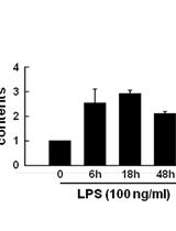
Mitochondria are essential regulators in not only ATP generation and metabolic reprogramming but also the generation of reactive oxygen species (ROS) in response to pathogenic stimuli. During exposure to environmental stresses including oxidative stress, exercise, cell division and caloric restriction, mitochondria can be divided to increase mitochondrial number, size, and mass. Moreover, mitochondrial biogenesis has a crucial role in the resolution of inflammation through preserving metabolic function. Recently, diverse biochemical methods have been utilized to evaluate activity of mitochondrial biogenesis. In this protocol, we will describe an in vitro assay to measure mitochondrial DNA content and mass. Quantitative real-time PCR analysis for determination of mitochondrial DNA content is a powerful tool with the addition of flow cytometry or confocal microscopy for evaluating mitochondrial mass. Together, these protocols may provide the significant information for mitochondria studies.

分子生物学

检测蛋白质相互作用的体外转录/翻译系统

An in vitro Transcription/translation System for Detection of Protein Interaction

检测蛋白质相互作用的体外转录/翻译系统

PL Pin-Chun Lin
YC Ya-Chun Chang
SL Shih-Shun Lin
16747 Views
May 5, 2016
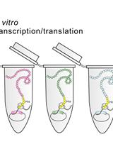
Studying protein-protein interaction is crucial to understand the fundamental processes of molecular biology. High-throughput screening, such as immunoprecipitation followed by proteomic analysis, allows for the identification of numerous candidate partners that might interact with a selected protein. However, experimental validation of protein-protein interaction requires conventional cloning and recombinant protein expression/purification, which are complicated and labor-intensive techniques. Here, we demonstrate an efficient experimental pipeline for verifying protein-protein interactions between a bait protein using the example of Odontoglossum ringspot virus (ORSV) capsid protein (CP) and the host CP-binding protein. These candidate CP-binding proteins were identified through high-throughput proteomic and transcriptomic approaches. Using the TOPO cloning strategy, each candidate gene was cloned into an expression vector for the expression of His-tagged recombinant proteins in a single step of an in vitro transcription/translation system. Such expressed His-tagged candidates can be used as prey with the CP bait protein in a co-immunoprecipitation (co-IP) assay to verify their physical interaction. Without the need for traditional protein expression and purification, this pipeline simplifies the validation process and provides a solution for high-throughput protein-protein interaction studies.

神经科学

啮齿动物社会认知记忆测试

Social Recognition Memory Test in Rodents

啮齿动物社会认知记忆测试

SJ Stephanie A. Jacobs
FH Fengying Huang
JT Joe Z. Tsien
WW Wei Wei
15898 Views
May 5, 2016
Social recognition memory is essential for the establishment and maintenance of a rodent colony. Recognition memory is important for social hierarchy, mate and offspring recognition, and interspecies recognition. Interspecies recognition is vital for recognizing frequent visitors to the animal’s habitat and whether or not the visitors pose a threat to the animals or colony (Macbeth et al., 2009; Noack et al., 2010). Here, we describe a protocol which effectively and reproducibly measures the social recognition for a juvenile male, a female, a mouse of another strain, and a rat. This task relies on the animal’s innate tendency to explore a novel social partner and decrease the exploration of a known familiar social partner (Thor et al., 1982). A significant decrease in the exploration of a partner from the training session to the recall session demonstrates a memory of the social partner. Also, we describe a social recognition procedure, the habituation-dishabituation paradigm that closely mimics typical short, frequent interactions between animals in a colony (Dantzer et al., 1987; Winslow and Camacho, 1995). Further, olfaction is a key component of social recognition, to test olfaction see Jacobs et al. (2016). In this protocol, we use transgenic NR2A overexpression mice to demonstrate how an impairment in social recognition memory may appear.
小鼠嗅觉认知记忆测试

Olfactory Recognition Memory Test in Mice

小鼠嗅觉认知记忆测试

SJ Stephanie A. Jacobs
FH Fengying Huang
JT Joe Z. Tsien
WW Wei Wei
9350 Views
May 5, 2016
Olfactory memory is an ethologically relevant task that relies on a mouse’s innate ability to use olfaction to forage for food (Zou et al., 2015), and identify safe foods. Although many of the same brain areas involved in other forms of memory are also involved in olfactory memory, the mechanisms are different (Sanchez-Andrade et al., 2005; Tong et al., 2014). Here, we describe one way to test olfactory memory in mice. The protocol described can be used to test long-term memory (memory which requires de novo protein synthesis) or short term memory by adjusting the delay time between the training session and the recall session (Freedman et al., 2013) and has been designed to mimic the single presentation of the social recognition paradigm. This paradigm relies on the mouse’s innate tendency to investigate a novel scent more than a familiar scent. Transgenic NR2A overexpression mice are known to have impaired long-term olfactory memory, but intact short-term memory, and are used here to demonstrate how one form of impaired olfactory memory may appear. Other genetically or chemically manipulated mice may be used in place of the transgenic mice used here.

植物科学

玉米胚芽鞘和主根组织中的内质网(ER)微粒体制备和IAA的定量测定

ER Microsome Preparation and Subsequent IAA Quantification in Maize Coleoptile and Primary Root Tissue

玉米胚芽鞘和主根组织中的内质网(ER)微粒体制备和IAA的定量测定

Verena Kriechbaumer Verena Kriechbaumer
10939 Views
May 5, 2016
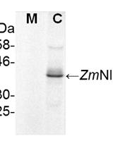
Auxin is a major growth hormone in plants and the first plant hormone to be discovered and studied (Darwin and Darwin, 1880). The auxin molecule in plants was first identified as indole-3-acetic acid (IAA) by Kögl et al. (1934). Active research over nearly a decade has shed light on many of the molecular mechanisms of its action but the complexity and redundancy of the auxin biosynthetic network raises questions about control of this system. We have shown that some enzymes involved in the YUCCA-route of auxin biosynthesis are not cytosolic but localised to the endoplasmic reticulum (ER) in both Arabidopsis thaliana (YUCCA4.2) (Kriechbaumer et al., 2012) as well as Zea mays (ZmTAR1 and ZmSPI) (Kriechbaumer et al., 2015). This is raising the intriguing possibility of subcellular compartmentation of auxin biosynthesis. To show that maize auxin biosynthesis indeed can take place in microsomal as well as cytosolic cellular fractions from maize seedlings we applied the protocol described here: Microsomes are being isolated from maize coleoptile and primary root tissue, enzyme assays with microsomal and cytosolic fractions using either tryptophan (Trp) or indole- -3pyruvic acid (IPyA) as a substrate are carried out and the auxin IAA is extracted and quantified.
拟南芥种子总粘液提取物中单糖的分析

Analysis of Monosaccharides in Total Mucilage Extractable from Arabidopsis Seeds

拟南芥种子总粘液提取物中单糖的分析

CV Cătălin Voiniciuc
MG Markus Günl
10634 Views
May 5, 2016
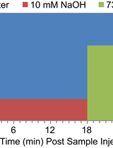
The Arabidopsis thaliana seed coat epidermis produces copious amounts of mucilage polysaccharides (Haughn and Western, 2012). Characterization of mucilage mutants has identified novel genes required for cell wall biosynthesis and modification (North et al., 2014). The biochemical analysis of seed mucilage is essential to evaluate how different mutations affect cell wall structure (Voiniciuc et al., 2015c). Here we describe a robust method to screen the monosaccharide composition of Arabidopsis seed mucilage using ion chromatography (IC). Mucilage from up to 48 samples can be extracted and prepared for IC analysis within 24 h (only 4 h hands-on). Furthermore, this protocol enables fast separation (31 min per sample), automatic detection and quantification of both neutral and acidic sugars.
拟南芥种子粘液释放的定量测定

Quantification of the Mucilage Detachment from Arabidopsis Seeds

拟南芥种子粘液释放的定量测定

CV Cătălin Voiniciuc
8202 Views
May 5, 2016
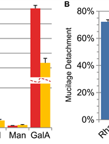
The Arabidopsis thaliana seed coat produces large amounts of cell wall polysaccharides, which swell out of the epidermal cells upon hydration of the mature dry seeds. While most mucilage polymers immediately diffuse in the surrounding solution, the remaining fraction tightly adheres to the seed, forming a dense gel-like capsule (Macquet et al., 2007). Recent evidence suggests that the adherence of mucilage is mediated by complex interactions between several cell wall components (Griffiths et al., 2014; Voiniciuc et al., 2015a). Therefore, it is important to evaluate how different cell wall mutants impact this mucilage property. This protocol facilitates the analysis of monosaccharides in sequentially extracted mucilage fractions, and quantifies the detachment of each component from seeds.

干细胞

从大鼠膝关节制备滑膜间充质干细胞

Preparation of Synovial Mesenchymal Stem Cells from a Rat Knee Joint

从大鼠膝关节制备滑膜间充质干细胞

Nobutake Ozeki Nobutake Ozeki
Takeshi Muneta Takeshi Muneta
Mitsuru Mizuno Mitsuru Mizuno
Ichiro Sekiya Ichiro Sekiya
9690 Views
May 5, 2016
Mesenchymal stem cells (MSCs), first described in human bone marrow, are emerging as promising cell-based therapeutics for a wide range of diseases (Caplan and Correa, 2011). MSCs have been isolated from various organs in the body, and synovial MSCs were first reported by De Bari et al. (2001). We previously reported that synovial MSCs have superior proliferation and chondrogenic potentials as compared to bone marrow-, muscle-, and adipose- derived MSCs in humans (Sakaguchi et al., 2005) and rats (Yoshimura et al., 2007). In addition, administration of synovial MSCs for osteochondral defect promoted cartilage regeneration in a rabbit (Koga et al., 2008) and a pig model (Nakamura et al., 2012). In 2008, we started a clinical trial in human and obtained satisfactory results of symptoms and regenerated cartilage by Magnetic Resonance Imaging (Sekiya et al., 2015). We have also engaged in multiple research lines using synovial MSCs for meniscus regeneration in rats (Horie et al., 2009; Horie et al., 2012; Katagiri et al., 2013; Okuno et al., 2014; Ozeki et al., 2015). In this article, we demonstrated how to harvest the synovium including infrapatellar fat pad from a rat knee joint, and to describe the technique of isolation and culture of rat synovial MSCs.