往期刊物2025

卷册: 15, 期号: 11

left

Oct

5

Sep

20

Sep

5

Aug

20

Aug

5

Jul

20

Jul

5

Jun

20

Jun

5

May

20

May

5

Apr

20

Apr

5

Mar

20

Mar

5

Feb

20

Feb

5

Jan

20

Jan

5

right

生物化学

源自出芽酵母的荧光蛋白融合PRPP转酰胺酶的体外冷凝实验

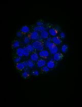
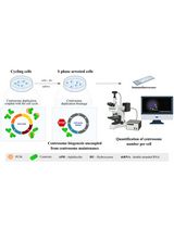
In vitro Condensation Assay of Fluorescent Protein-Fused PRPP Amidotransferase Purified from Budding Yeast Cells

源自出芽酵母的荧光蛋白融合PRPP转酰胺酶的体外冷凝实验

Masak Takaine Masak Takaine
980 Views
Jun 5, 2025
De novo synthesis of purine nucleotide is essential for the production of genetic materials and cellular chemical energy. PRPP amidotransferase (PPAT) is the rate-limiting enzyme in de novo purine synthesis, thereby playing a crucial regulatory role in this pathway. Recent studies suggest that metabolic enzymes, including PPAT, form condensates through phase separation to regulate cellular metabolism in response to environmental changes. However, due to the lack of methods for purifying eukaryotic PPAT, the biophysical properties of the enzyme have remained unknown. Here, I describe a protocol for purifying budding yeast PPAT tagged with green fluorescent protein from yeast cells, as well as an in vitro assay to examine condensation of the fluorescent PPAT by microscopy. These techniques enabled us to elucidate the mechanism controlling PPAT condensation and may also be applicable to the purification and condensation assay of other enzymes.

生物工程

在脱矿骨纸上共培养小鼠成骨细胞与破骨细胞用于研究骨重塑

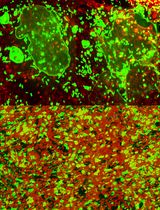
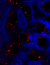
Murine Osteoblast and Osteoclast Co-culture on Demineralized Bone Paper for Bone Remodeling

在脱矿骨纸上共培养小鼠成骨细胞与破骨细胞用于研究骨重塑

SA Seema Amin
HY Hyejin Yoon
DC Dong-Hee Choi
YH Yi-Hao Hsu
JL Jungwoo Lee
1205 Views
Jun 5, 2025
Continuous and balanced bone remodeling is essential for maintaining mechanical integrity, mineral homeostasis, and hematopoiesis. Dysregulated bone metabolism develops pathological conditions, such as osteoporosis and bone metastasis. Functional and analytical recapitulation of bone remodeling in vitro is critical for advancing our understanding of bone mineral metabolism, disease mechanisms, and drug development. However, conventional models fail to replicate the essential complexity of the bone extracellular matrix (ECM) and the dynamic interplay between bone-forming osteoblasts and bone-resorbing osteoclasts. Recently, we developed an osteoid-mimicking demineralized bone paper (DBP) by thin-sectioning demineralized bovine compact bone matrix. DBP supports osteoblastic mineral deposition and the subsequent transition to bone-lining cells. When co-cultured with bone marrow mononuclear cells under biochemical stimulation, osteoblasts shift their regulatory secretion profiles and effectively induce osteoclastogenesis. The semi-transparent nature of DBP, combined with primary osteogenic cells retrieved from DsRed and eGFP reporter mice, enables longitudinal fluorescent monitoring of these multicellular processes and quantitative analysis. In this protocol, we describe the methods for DBP generation, reconstituting mineralized bone tissue complexity with osteoblasts, and recapitulating the bone remodeling cycle through bone marrow monocytes co-culture under biochemical stimulation, offering a useful platform for the related and broader research community.

细胞生物学

利用GFP报告蛋白在活细胞中成像PI(4,5)P₂

PI(4,5)P2 Imaging Using a GFP Reporter in Living Cells

利用GFP报告蛋白在活细胞中成像PI(4,5)P₂

MA Mariam Alkandari
CM Christopher R. McMaster
MT Mahtab Tavasoli
1124 Views
Jun 5, 2025
Phosphatidylinositol 4,5-bisphosphate [PI(4,5)P2] is a phospholipid enriched on the cytoplasmic leaflet of the plasma membrane, where it plays important roles in membrane trafficking and cytoskeletal dynamics through proteins that directly bind to it. PI(4,5)P2 can be metabolized to other phosphorylated forms of phosphatidylinositol to regulate numerous processes such as cell growth and development. PI(4,5)P2 can also be hydrolyzed to generate the second messengers diacylglycerol (DAG) and inositol triphosphate (IP3). Altered metabolism or mislocalization of PI(4,5)P2 can perturb one or more of its functions and contribute to disease states. Here, we present a protocol to visualize and quantify the localization of PI(4,5)P2 in live cells. The protocol uses a highly specific PI(4,5)P2 protein binding domain coupled to enhanced green fluorescence protein (PH-PLCD1-GFP), enabling localization and quantification of cytosol-facing PI(4,5)P2 to be determined. Localization and quantification of the PH-PLCD1-GFP, PI(4,5)P2 specific probe, is enabled by fluorescence imaging and confocal microscopy. This approach can be used to study the dynamics of PI(4,5)P2 localization temporally in live cells under both physiological and pathological conditions.
基于哺乳动物类器官球模型的内膜系统变化自动成像定量方法

An Automated Imaging Method for Quantification of Changes to the Endomembrane System in Mammalian Spheroid Models

基于哺乳动物类器官球模型的内膜系统变化自动成像定量方法

Margaritha M. Mysior Margaritha M. Mysior
JS Jeremy C. Simpson
977 Views
Jun 5, 2025
Three-dimensional cell models, such as spheroids, represent a more physiological arrangement in which cells can grow, allowing them to develop cell–cell interactions in all dimensions. The most common methods for growing spheroids are scaffold-based, typically using either extracellular matrix or hydrogels as a physical support for the cellular assembly. One key problem with this approach is that the spheroids that are produced can be highly variable in size and shape. The protocol presented here allows for the systematic production of uniform spheroids in a short time frame by utilising a micropatterned plate. We show that spheroids can be used to investigate fundamental research questions, such as how the endomembrane system is organised in cells. Our protocol can be used in a manual or automated manner, potentially allowing scaling up for screening applications. Furthermore, without the complication of removing the spheroids from the extracellular matrix or hydrogel, as would be required in scaffold-based systems, spheroids can easily be used in other downstream applications.
利用Seahorse XFe96通量分析仪检测PBMC的线粒体应激反应,并比较Poly-D-Lysine与Poly-L-Lysine的细胞附着性能

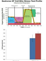
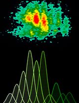
Mito Stress Assay of PBMCs With Seahorse XFe96 Flux Analyzer and Comparison of Poly-D-Lysine and Poly-L-Lysine for Cell Affinity

利用Seahorse XFe96通量分析仪检测PBMC的线粒体应激反应,并比较Poly-D-Lysine与Poly-L-Lysine的细胞附着性能

KS Kumudu Subasinghe
RB Raymond Berry III
MR Megan Rowe
AW Ali Winters
SY Shaohua Yang
NP Nicole Phillips
920 Views
Jun 5, 2025
The Seahorse 96 XF Analyzer (Agilent Technologies, Santa Clara, CA, USA) has been an effective tool in non-invasively measuring mitochondrial function for the past decade. It is a high-throughput respirometer that is considered the “gold standard” for quantifying mitochondrial function and bioenergetics in cells. Peripheral blood mononuclear cells (PBMCs) play a selective role in immune system responses and are key components of human immunity. Recent studies have suggested that these cell populations provide an overview of systemic changes within the body and therefore provide a source of sensitive biomarkers. Assessing mitochondrial function in PBMCs has been shown to provide an indication of metabolic stress associated with diseases such as diabetes and neurodegenerative conditions such as Alzheimer’s disease. In this protocol, we use two adhesive compounds, Poly-D-Lysine (PDL) and Poly-L-Lysine (PLL), at 50 μg/mL each per well, to immobilize PBMCs to a specialized Seahorse microplate to perform mitochondrial stress assay using the Seahorse Analyzer. We compared six cell densities of PBMCs to identify the optimal cell density for use in Seahorse Mito Stress analysis. This protocol includes the immobilization of freshly isolated PBM cells into a Seahorse microplate, hydration and calibration of the sensor cartridge, cell seeding, running the Seahorse Analyzer for the Mito Stress test, and simple data analysis to compare the effectiveness of PLL and PDL as the coating agent for PBMCs. The data analysis indicates that there is no statistical difference between PLL and PDL.

发育生物学

中心粒稳定性检测:研究果蝇培养细胞中中心体结构维持机制的方法

The Centriole Stability Assay: A Method to Investigate Mechanisms Involved in the Maintenance of the Centrosome Structure in Drosophila Cultured Cells

中心粒稳定性检测:研究果蝇培养细胞中中心体结构维持机制的方法

ML Mariana Lince-Faria
AF Ana Ferreira-Silva
AP Ana Pimenta-Marques
1342 Views
Jun 5, 2025
Centrosomes are vital eukaryotic organelles involved in regulating cell adhesion, polarity, mobility, and microtubule (MT) spindle assembly during mitosis. Composed of two centrioles surrounded by the pericentriolar material (PCM), centrosomes serve as the primary microtubule-organizing centers (MTOCs) in proliferating cells. The PCM is crucial for MT nucleation and centriole biogenesis. Centrosome numbers are tightly regulated, typically duplicating once per cell cycle, during the S phase. Deregulation of centrosome components can lead to severe diseases. While traditionally viewed as stable structures, centrosomes can be inactivated or disappear in differentiating cells, such as epithelial cells, muscle cells, neurons, and oocytes. Despite advances in understanding centrosome biogenesis and function, the mechanisms maintaining mature centrosomes or centrioles, as well as the pathways regulating their inactivation or elimination, remain less explored. Studying centrosome maintenance is challenging as it requires the uncoupling of centrosome biogenesis from maintenance. Tools for acute spatial-temporal manipulation are often unavailable, and manipulating multiple components in vivo is complex and time-consuming. This study presents a protocol that decouples centrosome biogenesis from maintenance, allowing the study of critical factors and pathways involved in the maintenance of the integrity of these important cellular structures.

环境生物学

自制可调组分纳米塑料及其应用:评估其对大鼠肺泡上皮细胞单层的生物电变化影响

In-house Fabrication of Nanoplastics of Tunable Composition and Application: Assessment of Bioelectric Changes in Primary Rat Lung Alveolar Epithelial Cell Monolayers Exposed to Nanoplastics

自制可调组分纳米塑料及其应用:评估其对大鼠肺泡上皮细胞单层的生物电变化影响

RC Ricki Chairil
JA Juan R. Alvarez
AS Arnold Sipos
NM Noah Malmstadt
EC Edward D. Crandall
KK Kwang-Jin Kim
1464 Views
Jun 5, 2025
Plastic pollution presents a looming danger to the environment and virtually all life on planet Earth. Especially pernicious are nanoplastics (NPs), which are plastic fragments with dimensions ≤1 μm. Conventional detection methods are ineffective for NPs, while their high specific surface area renders them efficient carriers of toxic substances; additionally, they may even be inherently toxic. Although NP waste chiefly arises from environmental weathering of larger plastic fragments, most published studies employed manufactured pristine NPs of uniform size and shape. Furthermore, almost all NP effects were studied using polystyrene (PS) as a convenient model material, despite PS accounting for 3 gas exposure to more closely mimic real environmental NPs. We also illustrate a simple and straightforward bioelectrical method for assessing passive and active ion transport properties of primary rat lung alveolar epithelial cell monolayers as a model for the distal mammalian lung exposed to one of the generated NPs. This protocol allows researchers to rapidly and more accurately assess the biological impact of various simulated environmental NPs on a vulnerable air–blood barrier in the lung.

免疫学

使用康可藻红素刺激冷冻保存的猪外周单个核细胞进行增殖检测,并结合FCS ExpressTM 7.18软件分析

Proliferation Assay Using Cryopreserved Porcine Peripheral Mononuclear Cells Stimulated With Concanavalin A and Analyzed With FCS ExpressTM 7.18 Software

使用康可藻红素刺激冷冻保存的猪外周单个核细胞进行增殖检测,并结合FCS ExpressTM 7.18软件分析

MB Marlene Bravo-Parra
RN Rahul K. Nelli
LY Lu Yen
Gino Castillo Gino Castillo
JM Juan Carlos Mora-Díaz
AC Ana F. Castillo-Espinoza
LG Luis G. Giménez-Lirola
1624 Views
Jun 5, 2025
In vitro lymphocyte proliferation assays are essential for assessing immune responses and antiproliferative drug efficacy. Such assays rely on antigen presentation or mitogen stimulation, with performance determined by reagent concentration and incubation time. Although splenocytes are often used, peripheral blood mononuclear cells (PBMCs) offer more accessible and practical sampling. However, a streamlined protocol for porcine PBMCs proliferation with robust batch analysis has been lacking. We therefore developed a detailed workflow for inducing proliferation in cryopreserved porcine PBMCs using 5 μg/mL concanavalin A (ConA). The protocol covers cell isolation, cryopreservation, ConA stimulation, CD4+ T-cell staining, flow cytometry acquisition and gating on an Attune NxT instrument, and batch analysis with FCS ExpressTM 7.18. This approach yielded 78.9% viable cells, of which 33.8% were CD4+ lymphocytes. Moreover, 93.9% (n = 216) of cells proliferated, yielding up to nine cell generations. Batch analysis in FCS ExpressTM enhanced the accuracy and interpretation of proliferation metrics. This validated protocol provides a reliable framework for generating consistent proliferation data in porcine immunology studies.

微生物学

利用壳聚糖纳米颗粒介导的dsRNA递送系统沉默丛枝菌根真菌基因

Silencing Arbuscular Mycorrhizal Fungal Gene Using Chitosan Nanoparticle-Mediated dsRNA Delivery System

利用壳聚糖纳米颗粒介导的dsRNA递送系统沉默丛枝菌根真菌基因

CY Chumei Yan
YW Yuemin Wang
QG Qianhuang Guo
HH Hongjuan Huan
SW Siwei Wang
XF Xiaoning Fan
XX Xianan Xie
1627 Views
Jun 5, 2025
It has been discovered that many phytopathogenic fungi can absorb exogenous double-stranded RNAs (dsRNAs) to silence target genes, inhibiting fungal growth and pathogenicity for plant protection. In our recent report, the beneficial arbuscular mycorrhizal (AM) fungi are capable of acquiring external naked dsRNAs; however, whether the dsRNAs can be delivered into AM fungi through nanocarriers remains to be investigated. Here, we introduce a simple and advanced method for in vitro synthesizing chitosan (CS)/dsRNA polyplex nanoparticles (PNs) to silence the target gene in the AM fungus Rhizophagus irregularis. This method is straightforward, requiring minimal modifications, and is both efficient and eco-friendly, offering potential for rapid application in elucidating gene functions in AM fungi.
iSLK细胞系的常规维护与KSHV再活化操作

General Maintenance and Reactivation of iSLK Cell Lines

iSLK细胞系的常规维护与KSHV再活化操作

Ariana C. Calderón-Zavala Ariana C. Calderón-Zavala
AM Aaron S. Mendez
Ekaterina E. Heldwein Ekaterina E. Heldwein
1185 Views
Jun 5, 2025
Since the establishment of the iSLK-BAC16 cell culture system, iSLK-BAC16 cells and their derivatives have been widely used for Kaposi’s sarcoma-associated herpesvirus (KSHV) studies. However, iSLK-BAC16 cells can be difficult to work with, in part due to the lack of standardized protocols and conflicting troubleshooting suggestions. Here, we describe the protocol for general iSLK-BAC16 cell culture and reactivation, which induces lytic KSHV replication and virion production. This protocol achieves robust levels of KSHV reactivation in our hands and can be readily used for studies of KSHV lytic infection mechanisms.

分子生物学

利用非还原性SDS-PAGE结合胰岛素原特异性免疫印迹定量分析折叠与错误折叠的胰岛素原形式

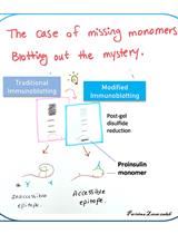
Quantification of Folded and Misfolded Proinsulin Forms Using Nonreducing SDS-PAGE and Proinsulin-Specific Immunoblotting

利用非还原性SDS-PAGE结合胰岛素原特异性免疫印迹定量分析折叠与错误折叠的胰岛素原形式

AA Anoop Arunagiri
IJ Insook Jang
PI Pamela Itkin-Ansari
RK Randal J. Kaufman
PA Peter Arvan
792 Views
Jun 5, 2025
We have observed that some proinsulin molecules in pancreatic islets and beta cell lines have incomplete or improper intramolecular disulfide bonds. These misfolded monomers can form intermolecular disulfide-linked complexes. Accurately quantifying the fraction of proinsulin molecules contained in these complexes is challenging. By proinsulin immunoblotting after nonreducing SDS-PAGE, the signal for disulfide-linked complexes can exceed the total proinsulin signal detected after reducing SDS-PAGE (i.e., overestimating the abundance of misfolded species due to antibody affinity differences). However, after modification of the SDS-PAGE and electrotransfer protocol, we have been able to more accurately estimate the fraction of proinsulin monomers folded to the native state, as well as misfolded proinsulin monomers and disulfide-linked complexes. Our improved technique offers the ability to obtain a more precise assessment of proinsulin misfolding under different environmental conditions in beta cells and normal islets and in diabetes.

神经科学

AMPA受体囊泡的单粒子追踪研究

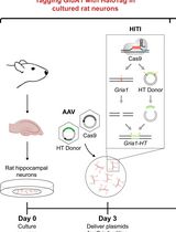
Single-Particle Tracking of AMPA Receptor-Containing Vesicles

AMPA受体囊泡的单粒子追踪研究

VW Victor C. Wong
DW Deepika Walpita
ZL Zhe J. Liu
EO Erin K. O’Shea
1249 Views
Jun 5, 2025
AMPA-type receptors are transported large distances to support synaptic plasticity at distal dendritic locations. Studying the motion of AMPA receptor+ vesicles can improve our understanding of the mechanisms that underlie learning and memory. Nevertheless, technical challenges that prevent the visualization of AMPA receptor+ vesicles limit our ability to study how these vesicles are trafficked. Existing methods rely on the overexpression of fluorescent protein-tagged AMPA receptors from plasmids, resulting in a saturated signal that obscures vesicles. Photobleaching must be applied to detect individual AMPA receptor+ vesicles, which may eliminate important vesicle populations from analysis. Here, we present a protocol to study AMPA receptor+ vesicles that addresses these challenges by 1) tagging AMPA receptors expressed from native loci with HaloTag and 2) employing a block-and-chase strategy with Janelia Fluor-conjugated HaloTag ligand to achieve sparse AMPA receptor labeling that obviates the need for photobleaching. After timelapse imaging is performed, AMPA receptor+ vesicles can be identified during image analysis, and their motion can be characterized using a single-particle tracking pipeline.
局部电泳给药诱导长期突触抑制的方法

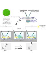
Local Iontophoretic Application for Pharmacological Induction of Long-Term Synaptic Depression

局部电泳给药诱导长期突触抑制的方法

Borys Olifirov Borys Olifirov
OF Oleksandra Fedchenko
AD Alexandr Dovgan
DB Daria Babets
VK Volodymyr Krotov
VC Volodymyr Cherkas
PB Pavel Belan
1012 Views
Jun 5, 2025
Long-term depression (LTD), a key form of synaptic plasticity, is typically induced through regulated Ca2+ entry via NMDA receptors and achieved by prolonged (up to hundreds of seconds) low-frequency presynaptic stimulation or bath application of NMDA receptor agonists. Electrophysiological approach to LTD induction requires specialized equipment, while bath applications limit productivity, as only one neuron per sample may be recorded. Here, we present a simple and effective protocol for pharmacological modeling of LTD in primary cultured neurons. This approach relies on highly localized iontophoretic application of NMDA, which induces LTD in individual cells, enhancing experimental throughput. We have analyzed spatio-temporal patterns of iontophoretic drug delivery and demonstrated how this technique may be combined with electrophysiological and live-cell imaging approaches to investigate LTD-related changes in synaptic strength and Ca2+-dependent signaling of neuronal Ca2+ sensor proteins.

植物科学

利用活细胞成像系统研究拟南芥珠被中淀粉体的复制

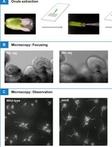
Using a Live Analysis System to Study Amyloplast Replication in Arabidopsis Ovule Integuments

利用活细胞成像系统研究拟南芥珠被中淀粉体的复制

MF Makoto T. Fujiwara
RA Rin Arakawa
TA Tomoko Abe
RI Ryuuichi D. Itoh
1653 Views
Jun 5, 2025
Amyloplasts, non-photosynthetic plastids specialized for starch synthesis and storage, proliferate in storage tissue cells of plants. To date, studies of amyloplast replication in roots and the ovule nucelli from various plant species have been performed using electron and fluorescence microscopy. However, a complete understanding of amyloplast replication remains unclear due to the absence of experimental systems capable of tracking their morphology and behavior in living cells. Recently, we demonstrated that Arabidopsis ovule integument could provide a platform for live-cell imaging of amyloplast replication. This system enables precise analysis of amyloplast number and shape, including the behavior of stroma-filled tubules (stromules), during proplastid-to-amyloplast development in post-mitotic cells. Here, we provide technical guidelines for observing and quantifying amyloplasts using conventional fluorescence microscopy in wild-type and several plastid-division mutants of Arabidopsis.
通过简并PCR鉴定二倍体马铃薯Solanum okadae中的S位点F-box蛋白序列

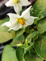
Identification of S-locus F-box Protein Sequences in Diploid Potato, Solanum okadae, via Degenerate PCR

通过简并PCR鉴定二倍体马铃薯Solanum okadae中的S位点F-box蛋白序列

AH Amar Hundare
1330 Views
Jun 5, 2025
In many plant species, self-incompatibility (SI) is a mechanism that inhibits inbreeding. SI is gametophytic in the Solanaceae, with specificity determined by S-ribonucleases (S-RNases) in the pistil and S-locus F-box proteins (SLFs) in the pollen. The role of these proteins has been studied extensively in the Solanaceae, often using Petunia as a model. Using degenerate PCR and Sanger sequencing, this protocol identified three SLF sequences from self-incompatible diploid potato (Solanum okadae). While SLFs are well-characterized in model species like Petunia, there is limited sequence data and no standardized protocols for identifying SLFs in non-model species such as S. okadae, hindering broader insights into SI across the Solanaceae. This protocol fills that gap by using degenerate PCR and Sanger sequencing with primers designed from conserved Petunia SLF regions to identify SLF sequences in S. okadae. SLF sequences from 10 distinct Solanaceae members sharing maximum identity with the S2-haplotype of Petunia were used to design two pairs of primers targeting different regions of the target sequence. PCR amplification using designed degenerate primers yielded amplicons that were directly sequenced and joined together to get the partial SLF sequence. It was observed that the S. okadae shared an orthologous relation with the Petunia SLF according to the phylogenetic analysis. These SLFs could be used in future SI breakdown experiments via the competitive interaction route. This protocol, including the primer design, is novel for detecting SLF sequences in S. okadae.