往期刊物2024

卷册: 14, 期号: 19

left

Dec

20

Dec

5

Nov

20

Nov

5

Oct

20

Oct

5

Sep

20

Sep

5

Aug

20

Aug

5

Jul

20

Jul

5

Jun

20

Jun

5

May

20

May

5

Apr

20

Apr

5

Mar

20

Mar

5

Feb

20

Feb

5

Jan

20

Jan

5

right

生物物理学

通过荧光寿命技术辅助探测具有亚细胞器分辨率的蛋白质聚集

Fluorescence Lifetime-Assisted Probing of Protein Aggregation with sub-Organellar Resolution

通过荧光寿命技术辅助探测具有亚细胞器分辨率的蛋白质聚集

KG Karnika Gupta
DM Daniel C. Maddison
EM Eduardo P. Melo
AD Ana Rosa M. da Costa
EA Edward Avezov
1090 Views
Oct 5, 2024
Protein misfolding fuels multiple neurodegenerative diseases, but existing techniques lack the resolution to pinpoint the location and physical properties of aggregates within living cells. Our protocol describes high-resolution confocal and fluorescent lifetime microscopy (Fast 3D FLIM) of an aggregation probing system. This system involves a metastable HaloTag protein (HT-aggr) labeled with P1 solvatochromic fluorophore, which can be targeted to subcellular compartments. This strategy allows to distinguish between aggregated and folded probe species, since P1 fluorophore changes its lifetime depending on the hydrophobicity of its microenvironment. The probe is not fluorescence intensity-dependent, overcoming issues related to intensity-based measurements of labeled proteins, such as control of probe quantity due to differences in expression or photobleaching of a proportion of the fluorophore population. Our approach reports on the performance of the machinery dealing with aggregation-prone substrates and thus opens doors to studying proteostasis and its role in neurodegenerative diseases.

细胞生物学

使用化学诱导二聚化系统快速调控内溶酶体上的磷脂酰肌醇磷酸盐

Acutely Modifying Phosphatidylinositol Phosphates on Endolysosomes Using Chemically Inducible Dimerization Systems

使用化学诱导二聚化系统快速调控内溶酶体上的磷脂酰肌醇磷酸盐

WY Wei Sheng Yap
PK Peter K. Kim
MB Maxime Boutry
905 Views
Oct 5, 2024
Phosphoinositides are rare membrane lipids that mediate cell signaling and membrane dynamics. PI(4)P and PI(3)P are two major phosphoinositides crucial for endolysosomal functions and dynamics, making them the lipids of interest in many studies. The acute modulation of phosphoinositides at a given organelle membrane can reveal important insights into their cellular function. Indeed, the localized depletion of PI(4)P and PI(3)P is a viable tool to assess the importance of these phosphoinositides in various experimental conditions. Here, we describe a live imaging method to acutely deplete PI(4)P and PI(3)P on endolysosomes. The depletion assay utilizes the GAI-GID1 or the FRB-FKBP inducible dimerization system to target the catalytic domain of the PI(4)P phosphatase, Sac1, or the PI(3)P phosphatase domain of MTM1 to the endolysosome for localized depletion of these phosphoinositides. By using the fluorescently tagged biosensors, 2xP4M and PX, we can validate and monitor the depletion of PI(4)P and PI(3)P, respectively, on endolysosomes in real-time. We discuss a method for normalizing the fluorescence measurements to appropriate the relative amount of these phosphoinositides in the organellar membranes (endolysosomes), which is required for monitoring PI(4)P or PI(3)P levels during the acute depletion assay. Since the localization of the dimerization partners is specified by the membrane targeting signal, our protocol will be useful for studying the signaling and functions of phosphoinositides at any membrane.
用于生物研究的水模型系统和细胞培养静态磁场暴露装置的构建与应用

Construction and Application of a Static Magnetic Field Exposure Apparatus for Biological Research in Aqueous Model Systems and Cell Culture

用于生物研究的水模型系统和细胞培养静态磁场暴露装置的构建与应用

JV Jana Vučković
HG Hakki Gurhan
BG Belen Gutierrez
JG Jose Guerra
LK Luke J. Kinsey
IN Iris Nava
AF Ashley Fitzpatrick
FB Frank S. Barnes
KT Kelly Ai-Sun Tseng
Wendy S. Beane Wendy S. Beane
834 Views
Oct 5, 2024
With the growth of the quantum biology field, the study of magnetic field (MF) effects on biological processes and their potential therapeutic applications has attracted much attention. However, most biologists lack the experience needed to construct an MF exposure apparatus on their own, no consensus standard exists for exposure methods, and protocols for model organisms are sorely lacking. We aim to provide those interested in entering the field with the ability to investigate static MF effects in their own research. This protocol covers how to design, build, calibrate, and operate a static MF exposure chamber (MagShield apparatus), with instructions on how to modify parameters to other specific needs. The MagShield apparatus is constructed of mu-metal (which blocks external MFs), allowing for the generation of experimentally controlled MFs via 3-axial Helmholtz coils. Precise manipulation of static field strengths across a physiologically relevant range is possible: nT hypomagnetic fields, μT to Xenopus, planarians, and fibroblast/fibrosarcoma cell lines, discussing the modifications needed for cell culture systems; however, the apparatus is easily adaptable to zebrafish, C. elegans, and 3D organoids. The operational methodology provided ensures uniform and reproducible results, affording the means for rigorous examination of static MF effects. Thus, this protocol is a valuable resource for investigators seeking to explore the intricate interplay between MFs and living organisms.

免疫学

在活细胞成像中测量趋化因子刺激下Jurkat细胞的Piezo1和肌动蛋白极性

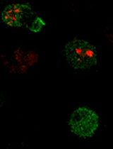
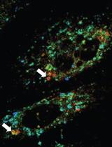
Measuring Piezo1 and Actin Polarity in Chemokine-Stimulated Jurkat Cells During Live-Cell Imaging

在活细胞成像中测量趋化因子刺激下Jurkat细胞的Piezo1和肌动蛋白极性

CL Chinky Shiu Chen Liu
PB Parijat Biswas
Dipyaman Ganguly Dipyaman Ganguly
922 Views
Oct 5, 2024
The process of T-lymphocyte migration involves a complex interplay of chemical and mechanical signals. Mechanotransduction mechanisms in T lymphocytes enable them to efficiently navigate through diverse architectural and topographical features of the dynamic tissue macro- and micro-niches encountered during immune responses. Piezo1 mechanosensors are crucial for driving optimal T-cell migration by driving actin-cytoskeletal remodeling. Chemokine-stimulated T lymphocytes demonstrate significant asymmetry or polarity of Piezo1 and actin along the cell axis. The establishment and maintenance of polarity in migrating cells are paramount for facilitating coordinated and directional movements along gradients of chemokine signals. Live-cell imaging techniques are widely employed to study the trajectories of migrating cells. Our approach expands upon current methodologies by not only tracking migrating cells but also imaging fluorescently labeled cellular components. Specifically, our method enables measurement of protein enrichment in the front and rear halves of the moving cell by analyzing the temporal direction of cell trajectories, subsequently bisecting the cell into front-back halves, and measuring the intensities of the fluorescent signals in each cell half at each time frame. Our protocol also facilitates the quantification of the angular distribution of fluorescent signals, enabling visualization of the spatial distribution of signals relative to the direction of cell migration. The protocol describes the examination of polarity in chemokine-treated Jurkat cells transfected with Piezo1-mCherry and actin-GFP constructs. This approach can be extended to live-cell imaging and polarity assessment of other fluorescently labeled proteins.

微生物学

使用iFAST在白色念珠菌中进行蛋白质的基因标记与成像

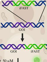
Genetic Tagging and Imaging of Proteins with iFAST in Candida albicans

使用iFAST在白色念珠菌中进行蛋白质的基因标记与成像

JD Jonas Devos
PV Patrick Van Dijck
WV Wouter Van Genechten
617 Views
Oct 5, 2024
Candida albicans is the most common human fungal pathogen, able to reside in a broad range of niches within the human body. Even though C. albicans systemic infection is associated with high mortality, the fungus has historically received relatively little attention, resulting in a lack of optimized molecular and fluorescent tools. Over the last decade, some extra focus has been put on the optimization of fluorescent proteins (FPs) of C. albicans. However, as the FPs are GFP-type, they require an aerobic environment and a relatively long period to fully mature. Recently, we have shown the application of a novel type of fluorogen-based FP, with an improved version of fluorescence activating and absorption shifting tag (iFAST), in C. albicans. Due to the dynamic relation between iFAST and its fluorogens, the system has the advantage of being reversible in terms of fluorescence. Furthermore, the combination of iFAST with different fluorogens results in different spectral and cellular properties, allowing customization of the system.

神经科学

使用免疫荧光技术对神经肌肉接头的可视化与分析

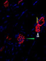
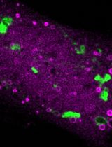
Visualization and Analysis of Neuromuscular Junctions Using Immunofluorescence

使用免疫荧光技术对神经肌肉接头的可视化与分析

YH You-Tian Hsieh
SC Show-Li Chen
1429 Views
Oct 5, 2024
The neuromuscular junction (NMJ) is an interface between motor neurons and skeletal muscle fibers, facilitating the transmission of signals that initiate muscle contraction. Its pivotal role lies in ensuring efficient communication between the nervous system and muscles, allowing for precise and coordinated movements essential for everyday activities and overall motor function. To provide insights into neuromuscular disease and development, understanding the physiology of NMJ is essential. We target acetylcholine receptors (AChR) by immunofluorescence assay (IFA) with α-bungarotoxin (BTX; snake venom neurotoxins binding to AChR) to visualize and quantify the NMJ. Changes in AChR distribution or structure can indicate alterations in receptor density, which may be associated with neuromuscular disorders or conditions that affect synaptic transmission. This protocol provides the methodology for isolating and longitudinally sectioning gastrocnemius muscle for AChR-targeted IFA for confocal microscopy and performing quantitative analysis of NMJs.

植物科学

高粱细胞外囊泡的分离、标记及相关光电显微技术

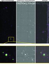
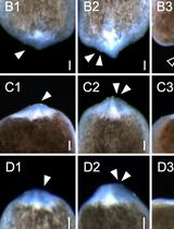
Sorghum bicolor Extracellular Vesicle Isolation, Labeling, and Correlative Light and Electron Microscopy

高粱细胞外囊泡的分离、标记及相关光电显微技术

DA Deji Adekanye
TC Timothy Chaya
JC Jeffrey L. Caplan
935 Views
Oct 5, 2024
Extracellular vesicles are membrane-bound organelles that play crucial roles in intercellular communication and elicit responses in the recipient cell, such as defense responses against pathogens. In this study, we have optimized a protocol for isolating extracellular vesicles (EVs) from Sorghum bicolor apoplastic wash. We characterized the EVs using fluorescence microscopy and correlative light and electron microscopy.

干细胞

获得人诱导多能干细胞系及三维培养的替代方法:一种简化且无需传代的低劳动量方法

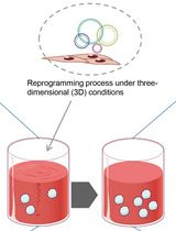
Alternative Method for Obtaining Human-Induced Pluripotent Stem Cell Lines and Three-Dimensional Growth: A Simplified, Passage-Free Approach that Minimizes Labor

获得人诱导多能干细胞系及三维培养的替代方法:一种简化且无需传代的低劳动量方法

MT Masaya Tsukamoto
TK Tomoyuki Kawasaki
AU Akihiro Umezawa
Hidenori Akutsu Hidenori Akutsu
1061 Views
Oct 5, 2024
Induced pluripotent stem cells (iPSCs) hold significant promise for numerous applications in regenerative medicine, disease modeling, and drug discovery. However, the conventional workflow for iPSC generation, with cells grown under two-dimensional conditions, presents several challenges, including the need for specialized scientific skills such as morphologically assessing and picking colonies and removing differentiated cells during the establishment phase. Furthermore, maintaining established iPSCs in three-dimensional culture systems, while offering scalability, necessitates an enzymatic dissociation step for their further growth in a complex and time-consuming protocol. In this study, we introduce a novel approach to address these challenges by reprogramming somatic cells grown under three-dimensional conditions as spheres using a bioreactor, thereby eliminating the need for two-dimensional culture and colony picking. The iPSCs generated in this study were maintained under three-dimensional conditions simply by transferring spheres to the next bioreactor, without the need for an enzymatic dissociation step. This streamlined method simplifies the workflow, reduces technical variability and labor, and paves the way for future advancements in iPSC research and its wider applications.