往期刊物2024

卷册: 14, 期号: 17

left

Dec

20

Dec

5

Nov

20

Nov

5

Oct

20

Oct

5

Sep

20

Sep

5

Aug

20

Aug

5

Jul

20

Jul

5

Jun

20

Jun

5

May

20

May

5

Apr

20

Apr

5

Mar

20

Mar

5

Feb

20

Feb

5

Jan

20

Jan

5

right

细胞生物学

FixNCut:单细胞检测样本可逆固定保存实用指南

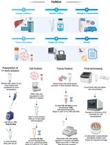
FixNCut: A Practical Guide to Sample Preservation by Reversible Fixation for Single Cell Assays

FixNCut:单细胞检测样本可逆固定保存实用指南

SW Shuoshuo Wang
LJ Laura Jiménez-Gracia
Ad Antonella Arruda de Amaral
IV Ioannis S. Vlachos
JP Jasmine Plummer
HH Holger Heyn
Luciano G. Martelotto Luciano G. Martelotto
2272 Views
Sep 5, 2024
The quality of standard single-cell experiments often depends on the immediate processing of cells or tissues post-harvest to preserve fragile and vulnerable cell populations, unless the samples are adequately fixed and stored. Despite the recent rise in popularity of probe-based and aldehyde-fixed RNA assays, these methods face limitations in species and target availability and are not suitable for immunoprofiling or assessing chromatin accessibility. Recently, a reversible fixation strategy known as FixNCut has been successfully deployed to separate sampling from downstream applications in a reproducible and robust manner, avoiding stress or necrosis-related artifacts. In this article, we present an optimized and robust practical guide to the FixNCut protocol to aid the end-to-end adaptation of this versatile method. This protocol not only decouples tissue or cell harvesting from single-cell assays but also enables a flexible and decentralized workflow that unlocks the potential for single-cell analysis as well as unconventional study designs that were previously considered unfeasible.

免疫学

用于测试抗PD-1和抗PD-L1抗体结合效力的基于成像的免疫突触PD-1定位检测方法

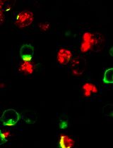
An Imaging-Based Assay to Measure the Location of PD-1 at the Immune Synapse for Testing the Binding Efficacy of Anti-PD-1 and Anti-PD-L1 Antibodies

用于测试抗PD-1和抗PD-L1抗体结合效力的基于成像的免疫突触PD-1定位检测方法

JZ Justin C. Zhong
SL Shalom Lerrer
AM Adam Mor
1486 Views
Sep 5, 2024
PD-1 is an immune checkpoint on T cells. Antibodies to PD-1 or its ligand PD-L1 are gaining popularity as a leading immunotherapy approach. In the US, 40% of all cancer patients will be treated with anti-PD-1 or anti-PD-L1 antibodies but, unfortunately, only 30% will respond, and many will develop immune-related adverse events. There are nine FDA-approved anti-PD-1/PD-L1 antibodies, and approximately 100 are in different stages of clinical development. It is a clinical challenge to choose the correct antibody for a given patient, and this is critical in advanced malignancies, which often do not permit a second-line intervention. To resolve that, an in vitro assay to compare the performance of the different anti-PD-1/PD-L1 antibodies is not only a critical tool for research purposes but also a possible tool for personalized medicine. There are some assays describing the binding affinity and function of anti-PD-1/PD-L1 antibodies. However, a significant limitation of existing assays is that they need to consider the location of PD-1 in the immune synapse, the interface between the T cell and tumor cells, and, therefore, ignore a critical component in its biology. To address this, we developed and validated an imaging-based assay to quantify and compare the ability of different anti-PD-1/PD-L1 antibodies to remove PD-1 from the immune synapse. We correlated that with the same antibodies' ability to increase cytokine secretion from the targeted cells. The strong correlation between PD-1 location and its function in vitro and in vivo within the antibody treatment setting validates this assay's usability, which is easily recordable and straightforward.

医学

内皮素-1诱导的鸡胚持续性缺血模型

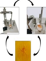
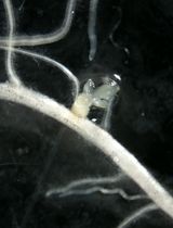
Endothelin-1-Induced Persistent Ischemia in a Chicken Embryo Model

内皮素-1诱导的鸡胚持续性缺血模型

NK Neha Kumari
RP Ravi Prakash
AS Abu J. Siddiqui
AW Arshi Waseem
MK Mohsin A. Khan
SR Syed S. Raza
938 Views
Sep 5, 2024
Current ischemic models strive to replicate ischemia-mediated injury. However, they face challenges such as inadequate reproducibility, difficulties in translating rodent findings to humans, and ethical, financial, and practical constraints that limit the accuracy of extensive research. This study introduces a novel approach to inducing persistent ischemia in 3-day-old chicken embryos using endothelin-1. The protocol targets the right vitelline arteries, validated with Doppler blood flow imaging and molecular biology experiments. This innovative approach facilitates the exploration of oxidative stress, inflammatory responses, cellular death, and potential drug screening suitability utilizing a 3-day-old chicken embryo.
血管性血友病因子活性检测的自动化预稀释系统

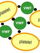
An Automated pre-Dilution Setup for Von Willebrand Factor Activity Assays

血管性血友病因子活性检测的自动化预稀释系统

TS Tobias Schachinger
AH Ann-Katrin Holik
GS Gerald Schrenk
HG Herbert Gritsch
SH Stefan Hofbauer
PF Paul G. Furtmüller
PT Peter L. Turecek
869 Views
Sep 5, 2024
Accurate quantification of von Willebrand factor ristocetin cofactor activity (VWF:RCo) is critical for the diagnosis and classification of von Willebrand disease, the most common hereditary and acquired bleeding disorder in humans. Moreover, it is important to accurately assess the function of von Willebrand factor (VWF) concentrates within the pharmaceutical industry to provide consistent and high-quality biopharmaceuticals. Although the performance of VWF:RCo assay has been improved by using coagulation analyzers, which are specialized devices for blood and blood plasma samples, scientists still report a high degree of intra- and inter-assay variation in clinical laboratories. Moreover, high, manual sample dilutions are required for VWF:RCo determination of VWF concentrates within the pharmaceutical industry, which are a major source for assay imprecision. For the first time, we present a precise and accurate method to determine VWF:RCo, where all critical pipetting and mixing steps are automated. A pre-dilution setup was established on CyBio FeliX (Analytik-Jena) liquid handling system, and an adapted VWF:RCo method on BCS-XP analyzer (Siemens) is used. The automated pre-dilution method was executed on three different, most frequently used coagulation analyzers and compared to manual pre-dilutions performed by an experienced operator. Comparative sample testing revealed a similar assay precision (coefficient of variation = 5.9% automated, 3.1% manual pre-dilution) and no significant differences between the automated approach and manual dilutions of an expert in this method. While no outliers were generated with the automated procedure, the manual pre-dilution resulted in an error rate of 8.3%. Overall, this operator-independent protocol enables standardization and offers an efficient way of fully automating VWF activity assays, while maintaining the precision and accuracy of an expert analyst.

微生物学

平行细菌趋化性检测的多重微流控平台

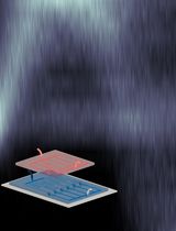
Multiplexed Microfluidic Platform for Parallel Bacterial Chemotaxis Assays

平行细菌趋化性检测的多重微流控平台

MS Michael R. Stehnach
RH Richard J. Henshaw
SF Sheri A. Floge
JG Jeffrey S. Guasto
1316 Views
Sep 5, 2024
The sensing of and response to ambient chemical gradients by microorganisms via chemotaxis regulates many microbial processes fundamental to ecosystem function, human health, and disease. Microfluidics has emerged as an indispensable tool for the study of microbial chemotaxis, enabling precise, robust, and reproducible control of spatiotemporal chemical conditions. Previous techniques include combining laminar flow patterning and stop-flow diffusion to produce quasi-steady chemical gradients to directly probe single-cell responses or loading micro-wells to entice and ensnare chemotactic bacteria in quasi-steady chemical conditions. Such microfluidic approaches exemplify a trade-off between high spatiotemporal resolution of cell behavior and high-throughput screening of concentration-specific chemotactic responses. However, both aspects are necessary to disentangle how a diverse range of chemical compounds and concentrations mediate microbial processes such as nutrient uptake, reproduction, and chemorepulsion from toxins. Here, we present a protocol for the multiplexed chemotaxis device (MCD), a parallelized microfluidic platform for efficient, high-throughput, and high-resolution chemotaxis screening of swimming microbes across a range of chemical concentrations. The first layer of the two-layer polydimethylsiloxane (PDMS) device comprises a serial dilution network designed to produce five logarithmically diluted chemostimulus concentrations plus a control from a single chemical solution input. Laminar flow in the second device layer brings a cell suspension and buffer solution into contact with the chemostimuli solutions in each of six separate chemotaxis assays, in which microbial responses are imaged simultaneously over time. The MCD is produced via standard photography and soft lithography techniques and provides robust, repeatable chemostimulus concentrations across each assay in the device. This microfluidic platform provides a chemotaxis assay that blends high-throughput screening approaches with single-cell resolution to achieve a more comprehensive understanding of chemotaxis-mediated microbial processes.

分子生物学

患者iPS细胞中C9orf72重复扩展的单分子测序

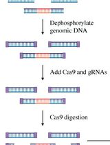
Single-Molecule Sequencing of the C9orf72 Repeat Expansion in Patient iPSCs

患者iPS细胞中C9orf72重复扩展的单分子测序

YT Yu-Chih Tsai
KB Katherine A. Brown
MB Mylinh T. Bernardi
JH John Harting
CC Claire D. Clelland
1428 Views
Sep 5, 2024
A hexanucleotide GGGGCC repeat expansion in the C9orf72 gene is the most frequent genetic cause of amyotrophic lateral sclerosis (ALS) and frontal temporal dementia (FTD). C9orf72 repeat expansions are currently identified with long-range PCR or Southern blot for clinical and research purposes, but these methods lack accuracy and sensitivity. The GC-rich and repetitive content of the region cannot be amplified by PCR, which leads traditional sequencing approaches to fail. We turned instead to PacBio single-molecule sequencing to detect and size the C9orf72 repeat expansion without amplification. We isolated high molecular weight genomic DNA from patient-derived iPSCs of varying repeat lengths and then excised the region containing the C9orf72 repeat expansion from naked DNA with a CRISPR/Cas9 system. We added adapters to the cut ends, capturing the target region for sequencing on PacBio’s Sequel, Sequel II, or Sequel IIe. This approach enriches the C9orf72 repeat region without amplification and allows the repeat expansion to be consistently and accurately sized, even for repeats in the thousands.

植物科学

评估土壤微生物组抑制高粱独脚金感染的机制

Evaluating Mechanisms of Soil Microbiome Suppression of Striga Infection in Sorghum

评估土壤微生物组抑制高粱独脚金感染的机制

TT Tamera Taylor
JD Jiregna Daksa
MS Mahdere Z. Shimels
DE Desalegn W. Etalo
BT Benjamin Thiombiano
AW Aimee Walmsey
AC Alexander J. Chen
HB Harro J. Bouwmeester
JR Jos M. Raaijmakers
SB Siobhan M. Brady
DK Dorota Kawa
1431 Views
Sep 5, 2024
The root parasitic weed Striga hermonthica has a devastating effect on sorghum and other cereal crops in Sub-Saharan Africa. Available Striga management strategies are rarely sufficient or not widely accessible or affordable. Identification of soil- or plant-associated microorganisms that interfere in the Striga infection cycle holds potential for development of complementary biological control measures. Such inoculants should be preferably based on microbes native to the regions of their application. We developed a method to assess microbiome-based soil suppressiveness to Striga with a minimal amount of field-collected soil. We previously used this method to identify the mechanisms of microbe-mediated suppression of Striga infection and to test individual microbial strains. Here, we present protocols to assess the functional potential of the soil microbiome and individual bacterial taxa that adversely affect Striga parasitism in sorghum via three major known suppression mechanisms. These methods can be further extended to other Striga hosts and other root parasitic weeds.
拟南芥子房中轴与侧区的激光辅助显微切割及高通量RNA测序

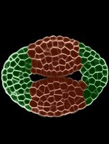
Laser-Assisted Microdissection and High-Throughput RNA Sequencing of the Arabidopsis Gynoecium Medial and Lateral Domains

拟南芥子房中轴与侧区的激光辅助显微切割及高通量RNA测序

VL Valentín Luna-García
Sd Stefan de Folter
1359 Views
Sep 5, 2024
For obtaining insights into gene networks during plant reproductive development, having transcriptomes of specific cells from developmental stages as starting points is very useful. During development, there is a balance between cell proliferation and differentiation, and many cell and tissue types are formed. While there is a wealth of transcriptome data available, it is mostly at the organ level and not at specific cell or tissue type level. Therefore, methods to isolate specific cell and tissue types are needed. One method is fluorescent activated cell sorting (FACS), but it has limitations such as requiring marker lines and protoplasting. Recently, single-cell/nuclei isolation methods have been developed; however, a minimum amount of genetic information (marker genes) is needed to annotate/predict the resulting cell clusters in these experiments. Another technique that has been known for some time is laser-assisted microdissection (LAM), where specific cells are microdissected and collected using a laser mounted on a microscope platform. This technique has advantages over the others because no fluorescent marker lines must be made, no marker genes must be known, and no protoplasting must be done. The LAM technique consists in tissue fixation, tissue embedding and sectioning using a microtome, microdissection and collection of the cells of interest on the microscope, and finally RNA extraction, library preparation, and RNA sequencing. In this protocol, we implement the use of normal slides instead of the membrane slides commonly used for LAM. We applied this protocol to obtain the transcriptomes of specific tissues during the development of the gynoecium of Arabidopsis.