往期刊物2024

卷册: 14, 期号: 1

left

Dec

20

Dec

5

Nov

20

Nov

5

Oct

20

Oct

5

Sep

20

Sep

5

Aug

20

Aug

5

Jul

20

Jul

5

Jun

20

Jun

5

May

20

May

5

Apr

20

Apr

5

Mar

20

Mar

5

Feb

20

Feb

5

Jan

20

Jan

5

right

生物化学

体外检测泪液对角膜上皮细胞功能的影响

In vitro Assay to Examine the Function of Tears on Corneal Epithelial Cells

体外检测泪液对角膜上皮细胞功能的影响

MM Moumita Mondal
MV Mehak Vohra
JS Jyoti Sangwan
SV Sudhir Verma
VC Vivien J. Coulson-Thomas
AC Arun Chandru
TB Tuhin Bhowmick
VS Virender S. Sangwan
MA Manisha Acharya
AT Anil Tiwari
1867 Views
Jan 5, 2024
Tears contain numerous secreted factors, enzymes, and proteins that help in maintaining the homeostatic condition of the eye and also protect it from the external environment. However, alterations to these enzymes and/or proteins during pathologies such as mechanical injury and viral or fungal infections can disrupt the normal ocular homeostasis, further contributing to disease development. Several tear film components have a significant role in curbing disease progression and promoting corneal regeneration. Additionally, several factors related to disease progression are secreted into the tear film, thereby serving as a valuable reservoir of biomarkers. Tears are readily available and can be collected via non-invasive techniques or simply from contact lenses. Tears can thus serve as a valuable and easy source for studying disease-specific biomarkers. Significant advancements have been made in recent years in the field of tear film proteomics, lipidomics, and transcriptomics to allow a better understanding of how tears can be utilized to gain insight into the etiology of diseases. These advancements have enabled us to study the pathophysiology of various disease states using tear samples. However, the mechanisms by which tears help to maintain corneal homeostasis and how they are able to form the first line of defense against pathogens remain poorly understood and warrant detailed in vitro studies. Herein, we have developed an in vitro assay to characterize the functional importance of patient isolated tears and their components on corneal epithelial cells. This novel approach closely mimics real physiological conditions and could help the researchers gain insight into the underlying mechanisms of ocular pathologies and develop new treatments.Key features• This method provides a new technique for analyzing the effect of tear components on human corneal epithelial cells.• The components of the tears that are altered in response to diseases can be used as a biomarker for detecting ocular complications.• This procedure can be further employed as an in vitro model for assessing the efficacy of drugs and discover potential therapeutic interventions.
使用荧光光谱法实时监测体外 ATG8 脂化

Real-Time Monitoring of ATG8 Lipidation in vitro Using Fluorescence Spectroscopy

使用荧光光谱法实时监测体外 ATG8 脂化

WZ Wenxin Zhang
TN Taki Nishimura
ST Sharon A. Tooze
1301 Views
Jan 5, 2024
Autophagy is an essential catabolic pathway used to sequester and engulf cytosolic substrates via a unique double-membrane structure, called an autophagosome. The ubiquitin-like ATG8 proteins play an important role in mediating autophagosome membrane expansion. They are covalently conjugated to phosphatidylethanolamine (PE) on the autophagosomes via a ubiquitin-like conjugation system called ATG8 lipidation. In vitro reconstitution of ATG8 lipidation with synthetic liposomes has been previously established and used widely to characterise the function of the E1 ATG7, the E2 ATG3, and the E3 complex ATG12–ATG5-ATG16L1. However, there is still a lack of a tool to provide kinetic measurements of this enzymatic reaction. In this protocol, we describe a real-time lipidation assay using NBD-labelled ATG8. This real-time assay can distinguish the formation of ATG8 intermediates (ATG7~ATG8 and/or ATG3~ATG8) and, finally, ATG8-PE conjugation. It allows kinetic characterisation of the activity of ATG7, ATG3, and the E3 complex during ATG8 lipidation. Furthermore, this protocol can be adapted to characterise the upstream regulators that may affect protein activity in ATG8 lipidation reaction with a kinetic readout.Key features• Preparation of ATG7 E1 from insect cells (Sf9 cells).• Preparation of ATG3 E2 from bacteria (E. coli).• Preparation of LC3B S3C from bacteria (E. coli).• Preparation of liposomes to monitor the kinetics of ATG8 lipidation in a real-time manner.Graphical overviewExperimental design to track the full reaction of ATG8 lipidation, described in this protocol

生物物理学

组合使用金属有机材料 (MOM)、电子顺磁共振 (EPR) 和质谱 (MS) 来描述蛋白水解过程的方案

A Protocol to Depict the Proteolytic Processes Using a Combination of Metal–Organic Materials (MOMs), Electron Paramagnetic Resonance (EPR), and Mass Spectrometry (MS)

组合使用金属有机材料 (MOM)、电子顺磁共振 (EPR) 和质谱 (MS) 来描述蛋白水解过程的方案

QL Qiaobin Li
ML Mary Lenertz
ZA Zoe Armstrong
AM Austin MacRae
LF Li Feng
AU Angel Ugrinov
ZY Zhongyu Yang
1123 Views
Jan 5, 2024
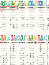
Proteolysis is a critical biochemical process yet a challenging field to study experimentally due to the self-degradation of a protease and the complex, dynamic degradation steps of a substrate. Mass spectrometry (MS) is the traditional way for proteolytic studies, yet it is challenging when time-resolved, step-by-step details of the degradation process are needed. We recently found a way to resolve the cleavage site, preference/selectivity of cleavage regions, and proteolytic kinetics by combining site-directed spin labeling (SDSL) of protein substrate, time-resolved two-dimensional (2D) electron paramagnetic resonance (EPR) spectroscopy, protease immobilization via metal–organic materials (MOMs), and MS. The method has been demonstrated on a model substrate and protease, yet there is a lack of details on the practical operations to carry out our strategy. Thus, this protocol summarizes the key steps and considerations when carrying out the EPR/MS study on proteolytic processes, which can be generalized to study other protein/polypeptide substrates in proteolysis. Details for the experimental operation and cautions of each step are reported with figures illustrating the concepts. This protocol provides an effective approach to understanding the proteolytic process with the advantages of offering time-resolved, residue-level resolution of structural basis underlying the process. Such information is important for revealing the cleavage site and proteolytic mechanisms of unknown proteases. The advantage of EPR, probing the target substrate regardless of the complexities caused by the proteases and their self-degradation, offers a practically effective, rapid, and easy-to-operate approach to studying proteolysis.Key features• Combining protease immobilization, EPR, spin labeling, and MS experimental methods allows for the analysis of proteolysis process in real time.• Reveals cleavage site, kinetics of product generation, and preference of cleavage regions via time-resolved SDSL-EPR.• MS confirms EPR findings and helps depict the sequences and populations of the cleaved segments in real time.• The demonstrated method can be generalized to other proteins or polypeptide substrates upon proteolysis by other proteases.Graphical overview

免疫学

外周血单核细胞体外扩增和极化人 γδT17 细胞

Expansion and Polarization of Human γδT17 Cells in vitro from Peripheral Blood Mononuclear Cells

外周血单核细胞体外扩增和极化人 γδT17 细胞

XC Xu Chen
XH Xiaoling Hu
FC Fuxiang Chen
JY Jun Yan
1374 Views
Jan 5, 2024
γδ T cells play a critical role in homeostasis and diseases such as infectious diseases and tumors in both mice and humans. They can be categorized into two main functional subsets: IFN-γ-producing γδT1 cells and IL-17-producing γδT17 cells. While CD27 expression segregates these two subsets in mice, little is known about human γδT17 cell differentiation and expansion. Previous studies have identified γδT17 cells in human skin and mucosal tissues, including the oral cavity and colon. However, human γδ T cells from peripheral blood mononuclear cells (PBMCs) primarily produce IFN-γ. In this protocol, we describe a method for in vitro expansion and polarization of human γδT17 cells from PBMCs.Key Features• Expansion of γδ T cells from peripheral blood mononuclear cells.• Human IL-17A-producing γδ T-cell differentiation and expansion using IL-7 and anti-γδTCR.• Analysis of IL-17A production post γδ T-cell expansion.

微生物学

重组酶聚合酶扩增-测流层析试纸 (RPA-LFD) 作为尖孢镰刀菌快速现场检测技术的开发

Development of Recombinase Polymerase Amplification–Lateral Flow Dipstick (RPA–LFD) as a Rapid On-Site Detection Technique for Fusarium oxysporum

重组酶聚合酶扩增-测流层析试纸 (RPA-LFD) 作为尖孢镰刀菌快速现场检测技术的开发

SH Shuodan Hu
HY Hong Yu
CZ Chuanqing Zhang
1335 Views
Jan 5, 2024
Fusarium oxysporum can cause many important plant diseases worldwide, such as crown rot, wilt, and root rot. During the development of strawberry crown rot, this pathogenic fungus spreads from the mother plant to the strawberry seedling through the stolon, with obvious characteristics of latent infection. Therefore, the rapid and timely detection of F. oxysporum can significantly help achieve effective disease management. Here, we present a protocol for the recombinase polymerase amplification– lateral flow dipstick (RPA–LFD) detection technique for the rapid detection of F. oxysporum on strawberry, which only takes half an hour. A significant advantage of our RPA–LFD technique is the elimination of the involvement of professional teams and laboratories, which qualifies it for field detection. We test this protocol directly on plant samples with suspected infection by F. oxysporum in the field and greenhouse. It is worth noting that this protocol can quickly, sensitively, and specifically detect F. oxysporum in soils and plants including strawberry.Key features• This protocol is used to detect whether plants such as strawberry are infected with F. oxysporum.• This protocol has potential for application in portable nucleic acid detection.• It can complete the detection of samples in the field within 30 min.Graphical overview
在人类永生化肌原 KD3 细胞中生成成熟的刚地弓形虫裂殖体

Generation of Mature Toxoplasma gondii Bradyzoites in Human Immortalized Myogenic KD3 Cells

在人类永生化肌原 KD3 细胞中生成成熟的刚地弓形虫裂殖体

DM Deborah Maus
BC Blake Curtis
DW David Warschkau
EB Estefanía Delgado Betancourt
FS Frank Seeber
MB Martin Blume
1329 Views
Jan 5, 2024
Toxoplasma gondii is a zoonotic protozoan parasite and one of the most successful foodborne pathogens. Upon infection and dissemination, the parasites convert into the persisting, chronic form called bradyzoites, which reside within cysts in muscle and brain tissue. Despite their importance, bradyzoites remain difficult to investigate directly, owing to limited in vitro models. In addition, the need for new drugs targeting the chronic stage, which is underlined by the lack of eradicating treatment options, remains difficult to address since in vitro access to drug-tolerant bradyzoites remains limited. We recently published the use of a human myotube-based bradyzoite cell culture system and demonstrated its applicability to investigate the biology of T. gondii bradyzoites. Encysted parasites can be functionally matured during long-term cultivation in these immortalized cells and possess many in vivo–like features, including pepsin resistance, oral infectivity, and antifolate resistance. In addition, the system is scalable, enabling experimental approaches that rely on large numbers, such as metabolomics. In short, we detail the cultivation of terminally differentiated human myotubes and their subsequent infection with tachyzoites, which then mature to encysted bradyzoites within four weeks at ambient CO2 levels. We also discuss critical aspects of the procedure and suggest improvements.Key features• This protocol describes a scalable human myotube-based in vitro system capable of generating encysted bradyzoites featuring in vivo hallmarks.• Bradyzoite differentiation is facilitated through CO2 depletion but without additional artificial stress factors like alkaline pH.• Functional maturation occurs over four weeks.Graphical overview

神经科学

微流体室中对区室化皮质神经元进行多重标记

Multiple Labeling of Compartmentalized Cortical Neurons in Microfluidic Chambers

微流体室中对区室化皮质神经元进行多重标记

GM Guillermo Moya-Alvarado
AA Alejandro Aguirre-Soto
FB Francisca C. Bronfman
1854 Views
Jan 5, 2024
Neurons are complex cells with two distinct compartments: the somatodendritic and the axonal domains. Because of their polarized morphology, it is challenging to study the differential cellular and molecular mechanisms that occur in axons and impact the soma and dendrites using conventional in vitro culture systems. Compartmentalized cultures offer a solution by physically and chemically separating the axonal from the somatodendritic domain of neurons. The microfluidic chamber model presented in this work is valuable for studying these mechanisms in primary cortical cultures derived from rat and mouse. In addition, this chamber model is compatible with various microscopy methods, such as phase contrast, and fluorescence imaging of living and fixed cells.Key features• Preparation and attachment of PDMS microfluidic chambers to glass coverslips.• Primary culture of cortical neurons and plating cortical neurons in microfluidic chamber.• Confirmation of compartmentalization using the retrograde transport of the fluorescently labeled form of cholera toxin subunit B (f-Ctb).• Immunofluorescence and multilabeling of compartmentalized cortical neurons.• Retrograde transport of fluorescently labeled BDNF.
设计 CRISPRoff 平台以调节骨髓细胞白血病(MCL-1)在定向少突胶质细胞神经前体细胞中的表达

Engineering a CRISPRoff Platform to Modulate Expression of Myeloid Cell Leukemia (MCL-1) in Committed Oligodendrocyte Neural Precursor Cells

设计 CRISPRoff 平台以调节骨髓细胞白血病(MCL-1)在定向少突胶质细胞神经前体细胞中的表达

MG Melanie Gil
CH Catherine A. Hamann
JB Jonathan M. Brunger
VG Vivian Gama
1588 Views
Jan 5, 2024
In vitro differentiation of human pluripotent stem cell (hPSC) model systems has furthered our understanding of human development. Techniques used to elucidate gene function during early development have encountered technical challenges, especially when targeting embryonic lethal genes. The introduction of CRISPRoff by Nuñez and collaborators provides an opportunity to heritably silence genes during long-term differentiation. We modified CRISPRoff and sgRNA Sleeping Beauty transposon vectors that depend on tetracycline-controlled transcriptional activation to silence the expression of embryonic lethal genes at different stages of differentiation in a stable manner. We provide instructions on how to generate sgRNA transposon vectors that can be used in combination with our CRISPRoff transposon vector and a stable hPSC line. We validate the use of this tool by silencing MCL-1, an anti-apoptotic protein, which results in pre-implantation embryonic lethality in mice; this protein is necessary for oligodendrocyte and hematopoietic stem cell development and is required for the in vitro survival of hPSCs. In this protocol, we use an adapted version of the differentiation protocol published by Douvaras and Fossati (2015) to generate oligodendrocyte lineage cells from human embryonic stem cells (hESCs). After introduction of the CRISPRoff and sgRNAs transposon vectors in hESCs, we silence MCL-1 in committed oligodendrocyte neural precursor cells and describe methods to measure its expression. With the methods described here, users can design sgRNA transposon vectors targeting MCL-1 or other essential genes of interest to study human oligodendrocyte development or other differentiation protocols that use hPSC model systems.Key features• Generation of an inducible CRISPRoff Sleeping Beauty transposon system.• Experiments performed in vitro for generation of inducible CRISPRoff pluripotent stem cell line amenable to oligodendrocyte differentiation.• Strategy to downregulate an essential gene at different stages of oligodendrocyte development.Graphical overviewWorkflow for generating inducible CRISPRoff stem cell line and assessing knockdown phenotype in stem cell–derived committed oligodendrocyte neural precursor cells