往期刊物2023
卷册: 13, 期号: 19
生物化学
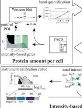
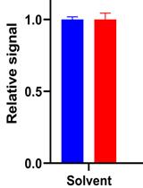
Protein Level Quantification Across Fluorescence-based Platforms
跨荧光平台的蛋白质水平定量
生物信息学与计算生物学
GutMap: A New Interface for Analysing Regional Motility Patterns in ex vivo Mouse Gastrointestinal Preparations
GutMap:用于分析离体小鼠胃肠道制剂中区域运动模式的新界面
生物科学
Production, Extraction, and Solubilization of Exopolysaccharides Using Submerged Cultures of Agaricomycetes
利用伞菌深层培养物生产、提取和溶解胞外多糖
细胞生物学
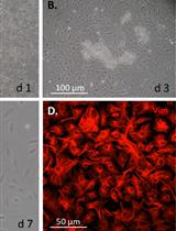
Mouse Corneal Epithelial and Stromal Cell Isolation and Culture
小鼠角膜上皮和基质细胞的分离和培养
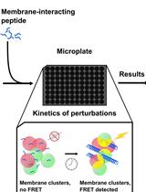
Fluorescence Resonance Energy Transfer to Detect Plasma Membrane Perturbations in Giant Plasma Membrane Vesicles
荧光共振能量转移检测巨型质膜囊泡中的质膜扰动
发育生物学
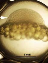
A Rapid and Simple Procedure for the Isolation of Embryonic Cells from Fish Eggs
一种快速简便的从鱼卵中分离胚胎细胞的方法
环境生物学
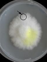
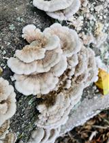
Co-culture Wood Block Decay Test with Bacteria and Wood Rotting Fungi to Analyse Synergism/Antagonism during Wood Degradation
细菌和木材腐烂真菌共培养木块腐烂实验,分析木材降解过程中的协同/拮抗作用
免疫学
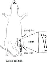
Isolation and Analysis of B-cell Progenitors from Bone Marrow by Flow Cytometry
通过流式细胞术分离和分析骨髓 B 细胞祖细胞
医学
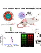
In vitro Quality Assessments of Perfluorocarbon Nanoemulsions for Near-infrared Fluorescence Imaging of Inflammation in Preclinical Models
用于临床前模型中炎症近红外荧光成像的全氟化碳纳米乳剂的体外质量评估
神经科学
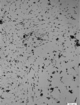
Isolation and Culture of Neural Stem/Progenitor Cells from the Hippocampal Dentate Gyrus of Young Adult and Aged Rats
青年和老年大鼠海马齿状回神经干/祖细胞的分离和培养
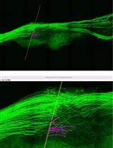
Application of Electrical Stimulation to Enhance Axon Regeneration Following Peripheral Nerve Injury
电刺激增强周围神经损伤后的轴突再生的应用
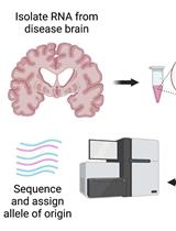
Testing for Allele-specific Expression from Human Brain Samples
测试人脑样本的等位基因特异性表达
植物科学
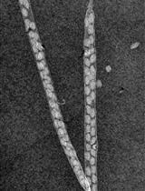
A Novel Imaging Protocol for Investigating Arabidopsis thaliana Siliques and Seeds Using X-rays
使用 X 射线研究拟南芥长角果和种子的新型成像方案