

往期刊物2015
卷册: 5, 期号: 22
癌症生物学
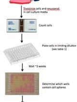
In vitro and in vivo Limiting Dilution Assay for Colorectal Cancer
结肠直肠癌的体外和体内限制稀释测定法
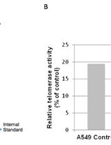
Telomerase Repeated Amplification Protocol (TRAP)
端粒酶重复扩增实验(TRAP)


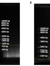
Telomere Restriction Fragment (TRF) Analysis
端粒限制酶片段(TRF)分析
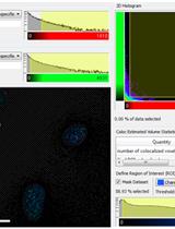
Telomere Dysfunction Induced Foci (TIF) Analysis
端粒功能失调诱导病灶(TIF)分析
13C Tracer Studies of Metabolism in Mouse Tumor Xenografts
13C 示踪研究老鼠肿瘤异种移植中的新陈代谢
免疫学
Skin Wound Healing Model - Excisional Wounding and Assessment of Lesion Area
皮肤伤口愈合模型——切除性创伤和病变评估


Estimation of Wound Tissue Neutrophil and Macrophage Accumulation by Measuring Myeloperoxidase (MPO) and N-Acetyl-β-D-glucosaminidase (NAG) Activities
通过测定髓过氧化物酶(MPO)和N-乙酰-β-D-氨基葡萄糖苷酶(NAG)的活性估计创伤组织的嗜中性白细胞和巨噬细胞的积聚
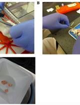
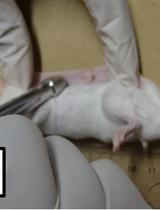
Permanent Occlusion of the Left Anterior Coronary Artery in the Rat
大鼠左前冠状动脉的永久性闭塞

微生物学
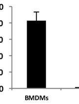
Purification of Bacterial RNA from Infected Macrophages
感染巨噬细胞的细菌RNA的纯化
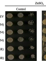
An Assay to Test the Capacity of Arabidopsis Plant Defensin Type1 Protein to Induce Cellular Zinc (Zn) Tolerance in Yeast
拟南芥防御素1型蛋白诱导酵母细胞中锌耐受性的能力的测试分析
神经科学
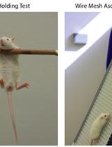
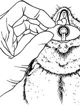
Locomotor Coordination Assay in Rats
大鼠中运动器官协调性的试验

植物科学
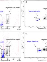
Sample Preparation and Fractionation of Arabidopsis thaliana Sperm and Vegetative Cell Nuclei by FACS
FACS法进行拟南芥精子和营养细胞核的样本制备和分离

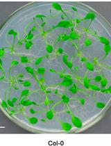
Arabidopsis Leaf Explant Culture
拟南芥叶外植体培养


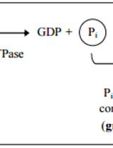
Expression, Purification and in vitro Enzyme Activity Assay of Plant Derived GTPase
植物源GTP酶的表达、纯化和体外酶活性的测定
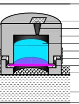
Quantifying the Permeability of the Apoplastic Water Barrier in Cosmos Petals
秋英属花瓣中质外体水屏障渗透性的定量测定