往期刊物2023

卷册: 13, 期号: 4

left

Dec

20

Dec

5

Nov

20

Nov

5

Oct

20

Oct

5

Sep

20

Sep

5

Aug

20

Aug

5

Jul

20

Jul

5

Jun

20

Jun

5

May

20

May

5

Apr

20

Apr

5

Mar

20

Mar

5

Feb

20

Feb

5

Jan

20

Jan

5

right

生物化学

使用 RNA 链霉亲和素适体标签分离 RNase MRP 的新方法

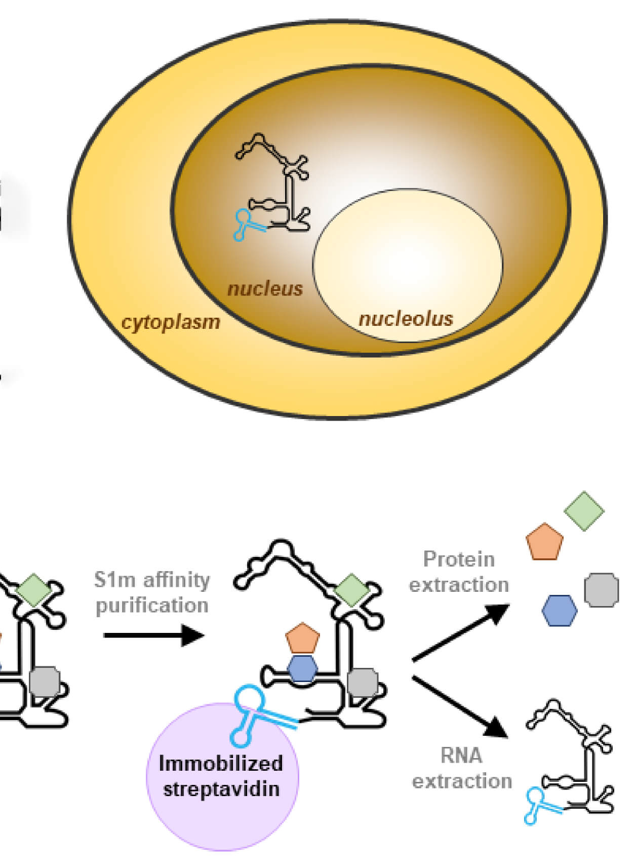
A Novel Method to Isolate RNase MRP Using RNA Streptavidin Aptamer Tags

使用 RNA 链霉亲和素适体标签分离 RNase MRP 的新方法

VC Violette Charteau
MD Merel Derksen
GP Ger J. M. Pruijn
1924 Views
Feb 20, 2023
Interactions between RNA-binding proteins and RNA molecules are at the center of multiple biological processes. Therefore, accurate characterization of the composition of ribonucleoprotein complexes (RNPs) is crucial. Ribonuclease (RNase) for mitochondrial RNA processing (MRP) and RNase P are highly similar RNPs that play distinct roles at the cellular level; as a consequence, the specific isolation of either of these complexes is essential to study their biochemical function. Since their protein components are nearly identical, purification of these endoribonucleases using protein-centric methods is not feasible. Here, we describe a procedure employing an optimized high-affinity streptavidin-binding RNA aptamer, termed S1m, to purify RNase MRP free of RNase P. This report details all steps from the RNA tagging to the characterization of the purified material. We show that using the S1m tag allows efficient isolation of active RNase MRP.
Far-western blotting 检测胰岛素受体底物与胰岛素受体的结合

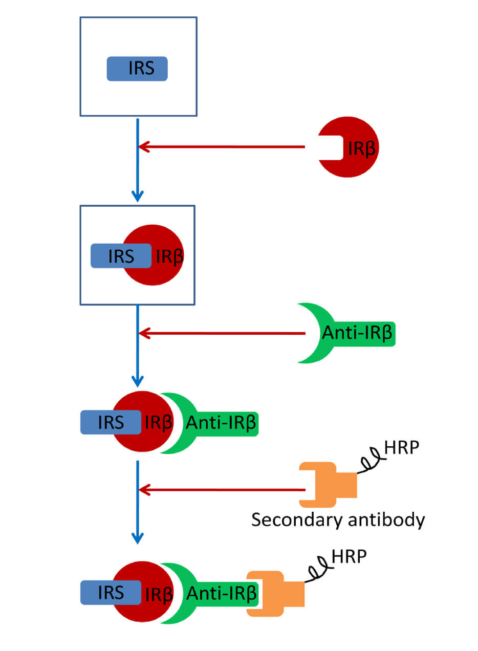
Far-western Blotting Detection of the Binding of Insulin Receptor Substrate to the Insulin Receptor

Far-western blotting 检测胰岛素受体底物与胰岛素受体的结合

JP Jinghua Peng
BR Balamurugan Ramatchandirin
AP Alexia Pearah
LH Ling He
1753 Views
Feb 20, 2023
Far-western blotting, derived from the western blot, has been used to detect interactions between proteins in vitro, such as receptor–ligand interactions. The insulin signaling pathway plays a critical role in the regulation of both metabolism and cell growth. The binding of the insulin receptor substrate (IRS) to the insulin receptor is essential for the propagation of downstream signaling after the activation of the insulin receptor by insulin. Here, we describe a step-by-step far-western blotting protocol for determining the binding of IRS to the insulin receptor.

生物科学

小鼠肌肉功能、瘦肌肉质量以及肌纤维分型的综合分析

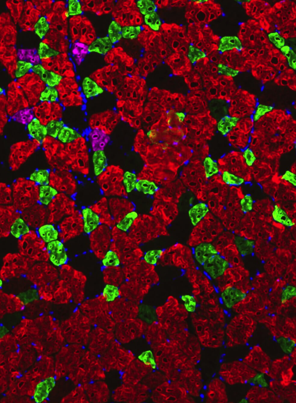
Comprehensive Analyses of Muscle Function, Lean and Muscle Mass, and Myofiber Typing in Mice

小鼠肌肉功能、瘦肌肉质量以及肌纤维分型的综合分析

HD Hima Bindu Durumutla
CV Chiara Villa
MP Manoj Panta
MW Michelle Wintzinger
AP Ashok Daniel Prabakaran
KM Karen Miz
MQ Mattia Quattrocelli
3481 Views
Feb 20, 2023
Skeletal muscle disorders commonly affect the function and integrity of muscles. Novel interventions bring new potential to rescue or alleviate the symptoms associated with these disorders. In vivo and in vitro testing in mouse models allows quantitative evaluation of the degree of muscle dysfunction, and therefore, the level of potential rescue/restoration by the target intervention. Several resources and methods are available to assess muscle function and lean and muscle mass, as well as myofiber typing as separate concepts; however, a technical resource unifying these methods is missing. Here, we provide detailed procedures for analyzing muscle function, lean and muscle mass, and myofiber typing in a comprehensive technical resource paper.Graphical abstract

生物物理学

在哺乳动物细胞中使用全内反射荧光显微镜 (TIRFM) 对膜蛋白进行成像

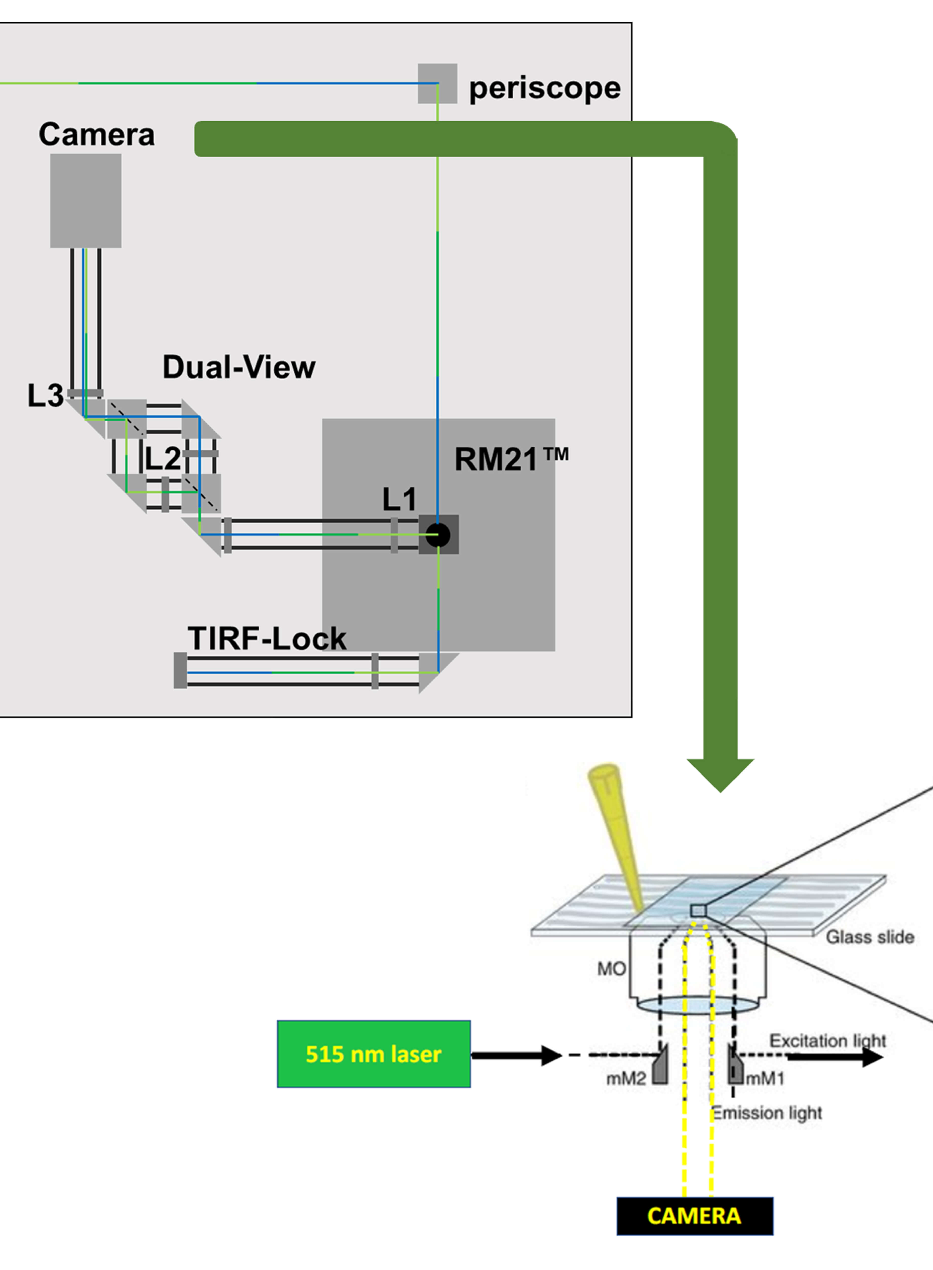
Imaging Membrane Proteins Using Total Internal Reflection Fluorescence Microscopy (TIRFM) in Mammalian Cells

在哺乳动物细胞中使用全内反射荧光显微镜 (TIRFM) 对膜蛋白进行成像

KG Kirin D. Gada
JK Jordie M. Kamuene
TK Takeharu Kawano
LP Leigh D. Plant
1343 Views
Feb 20, 2023
The cell surfaceome is of vital importance across physiology, developmental biology, and disease states alike. The precise identification of proteins and their regulatory mechanisms at the cell membrane has been challenging and is typically determined using confocal microscopy, two-photon microscopy, or total internal reflection fluorescence microscopy (TIRFM). Of these, TIRFM is the most precise, as it harnesses the generation of a spatially delimited evanescent wave at the interface of two surfaces with distinct refractive indices. The limited penetration of the evanescent wave illuminates a narrow specimen field, which facilitates the localization of fluorescently tagged proteins at the cell membrane but not inside of the cell. In addition to constraining the depth of the image, TIRFM also significantly enhances the signal-to-noise ratio, which is particularly valuable in the study of live cells. Here, we detail a protocol for micromirror TIRFM analysis of optogenetically activated protein kinase C-ϵ in HEK293-T cells, as well as data analysis to demonstrate the translocation of this construct to the cell-surface following optogenetic activation.Graphic abstract

细胞生物学

从新生小鼠心脏中分离和培养原代成纤维细胞以研究心脏纤维化

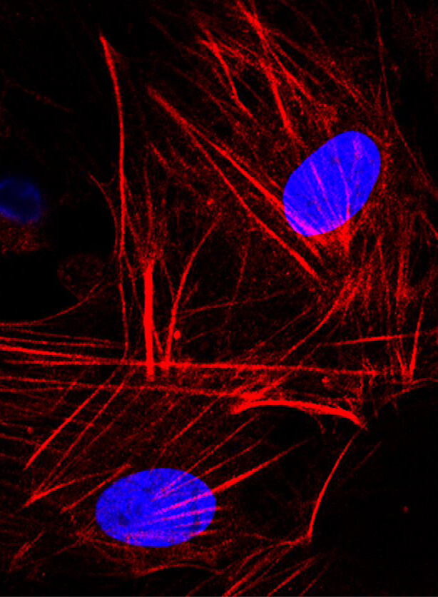
Isolation and Culture of Primary Fibroblasts from Neonatal Murine Hearts to Study Cardiac Fibrosis

从新生小鼠心脏中分离和培养原代成纤维细胞以研究心脏纤维化

SK Shweta Kumar
DN Dimple Nagesh
VR Venketsubbu Ramasubbu
AP Arathi Bangalore Prabhashankar
NS Nagalingam Ravi Sundaresan
2753 Views
Feb 20, 2023
Cardiac fibroblasts are one of the major constituents of a healthy heart. Cultured cardiac fibroblasts are a crucial resource for conducting studies on cardiac fibrosis. The existing methods for culturing cardiac fibroblasts involve complicated steps and require special reagents and instruments. The major problems faced with primary cardiac fibroblast culture are the low yield and viability of the cultured cells and contamination with other heart cell types, including cardiomyocytes, endothelial cells, and immune cells. Numerous parameters, including the quality of the reagents used for the culture, conditions maintained during digestion of the cardiac tissue, composition of the digestion mixture used, and age of the pups used for culture determine the yield and purity of the cultured cardiac fibroblasts. The present study describes a detailed and simplified protocol to isolate and culture primary cardiac fibroblasts from neonatal murine pups. We demonstrate the transdifferentiation of fibroblasts into myofibroblasts through transforming growth factor (TGF)-β1 treatment, representing the changes in fibroblasts during cardiac fibrosis. These cells can be used to study the various aspects of cardiac fibrosis, inflammation, fibroblast proliferation, and growth.

免疫学

杂交瘤细胞中基于 CRISPR/Cas9 的免疫球蛋白位点工程

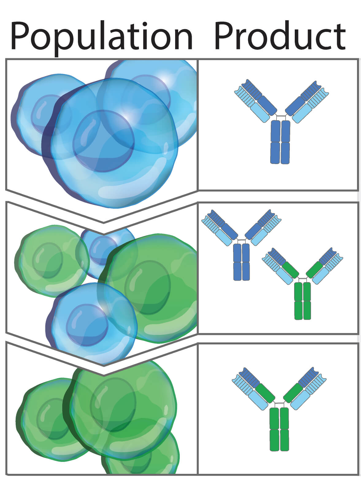
CRISPR/Cas9-based Engineering of Immunoglobulin Loci in Hybridoma Cells

杂交瘤细胞中基于 CRISPR/Cas9 的免疫球蛋白位点工程

CL Camille M. Le Gall
FF Felix L. Fennemann
Jv Johan M.S. van der Schoot
FS Ferenc A. Scheeren
MV Martijn Verdoes
1907 Views
Feb 20, 2023
Development of the hybridoma technology by Köhler and Milstein (1975) has revolutionized the immunological field by enabling routine use of monoclonal antibodies (mAbs) in research and development efforts, resulting in their successful application in the clinic today. While recombinant good manufacturing practices production technologies are required to produce clinical grade mAbs, academic laboratories and biotechnology companies still rely on the original hybridoma lines to stably and effortlessly produce high antibody yields at a modest price. In our own work, we were confronted with a major issue when using hybridoma-derived mAbs: there was no control over the antibody format that was produced, a flexibility that recombinant production does allow. We set out to remove this hurdle by genetically engineering antibodies directly in the immunoglobulin (Ig) locus of hybridoma cells. We used clustered regularly interspaced short palindromic repeats (CRISPR)/CRISPR-associated protein 9 (Cas9) and homology-directed repair (HDR) to modify antibody’s format [mAb or antigen-binding fragment (Fab’)] and isotype. This protocol describes a straightforward approach, with little hands-on time, leading to stable cell lines secreting high levels of engineered antibodies. Parental hybridoma cells are maintained in culture, transfected with a guide RNA (gRNA) targeting the site of interest in the Ig locus and an HDR template to knock in the desired insert and an antibiotic resistance gene. By applying antibiotic pressure, resistant clones are expanded and characterized at the genetic and protein level for their ability to produce modified mAbs instead of the parental protein. Finally, the modified antibody is characterized in functional assays. To demonstrate the versatility of our strategy, we illustrate this protocol with examples where we have (i) exchanged the constant heavy region of the antibody, creating chimeric mAb of a novel isotype, (ii) truncated the antibody to create an antigenic peptide-fused Fab’ fragment to produce a dendritic cell–targeted vaccine, and (iii) modified both the constant heavy (CH)1 domain of the heavy chain (HC) and the constant kappa (Cκ) light chain (LC) to introduce site-selective modification tags for further derivatization of the purified protein. Only standard laboratory equipment is required, which facilitates its application across various labs. We hope that this protocol will further disseminate our technology and help other researchers.Graphical abstract

神经科学

在小鼠中使用振动平台自动睡眠剥夺设置

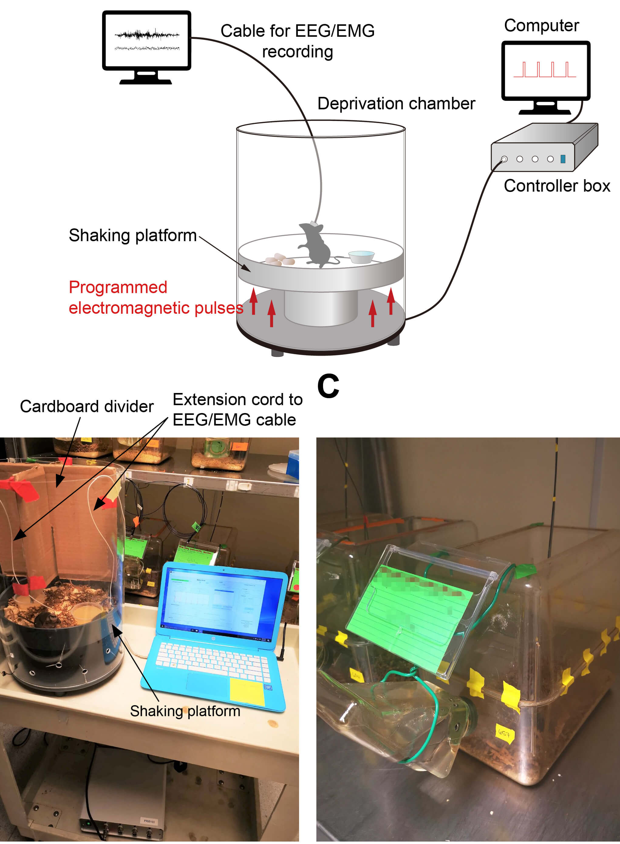
Automated Sleep Deprivation Setup Using a Shaking Platform in Mice

在小鼠中使用振动平台自动睡眠剥夺设置

WB Wen-Jie Bian
Ld Luis de Lecea
1424 Views
Feb 20, 2023
The functions of sleep remain largely unclear, and even less is known about its role in development. A general strategy to tackle these questions is to disrupt sleep and measure the outcomes. However, some existing sleep deprivation methods may not be suitable for studying the effects of chronic sleep disruption, due to their lack of effectiveness and/or robustness, substantial stress caused by the deprivation method, or consuming a large quantity of time and manpower. More problems may be encountered when applying these existing protocols to young, developing animals, because of their likely heightened vulnerability to stressors, and difficulties in precisely monitoring sleep at young ages. Here, we report a protocol of automated sleep disruption in mice using a commercially available, shaking platform–based deprivation system. We show that this protocol effectively and robustly deprives both non-rapid-eye-movement (NREM) sleep and rapid-eye-movement (REM) sleep without causing a significant stress response, and does not require human supervision. This protocol uses adolescent mice, but the method also works with adult mice.Graphical abstractAutomated sleep deprivation system. The platform of the deprivation chamber was programmed to shake in a given frequency and intensity to keep the animal awake while its brain and muscle activities were continuously monitored by electroencephalography and electromyography.
红外印迹法检测斑马鱼视网膜蛋白

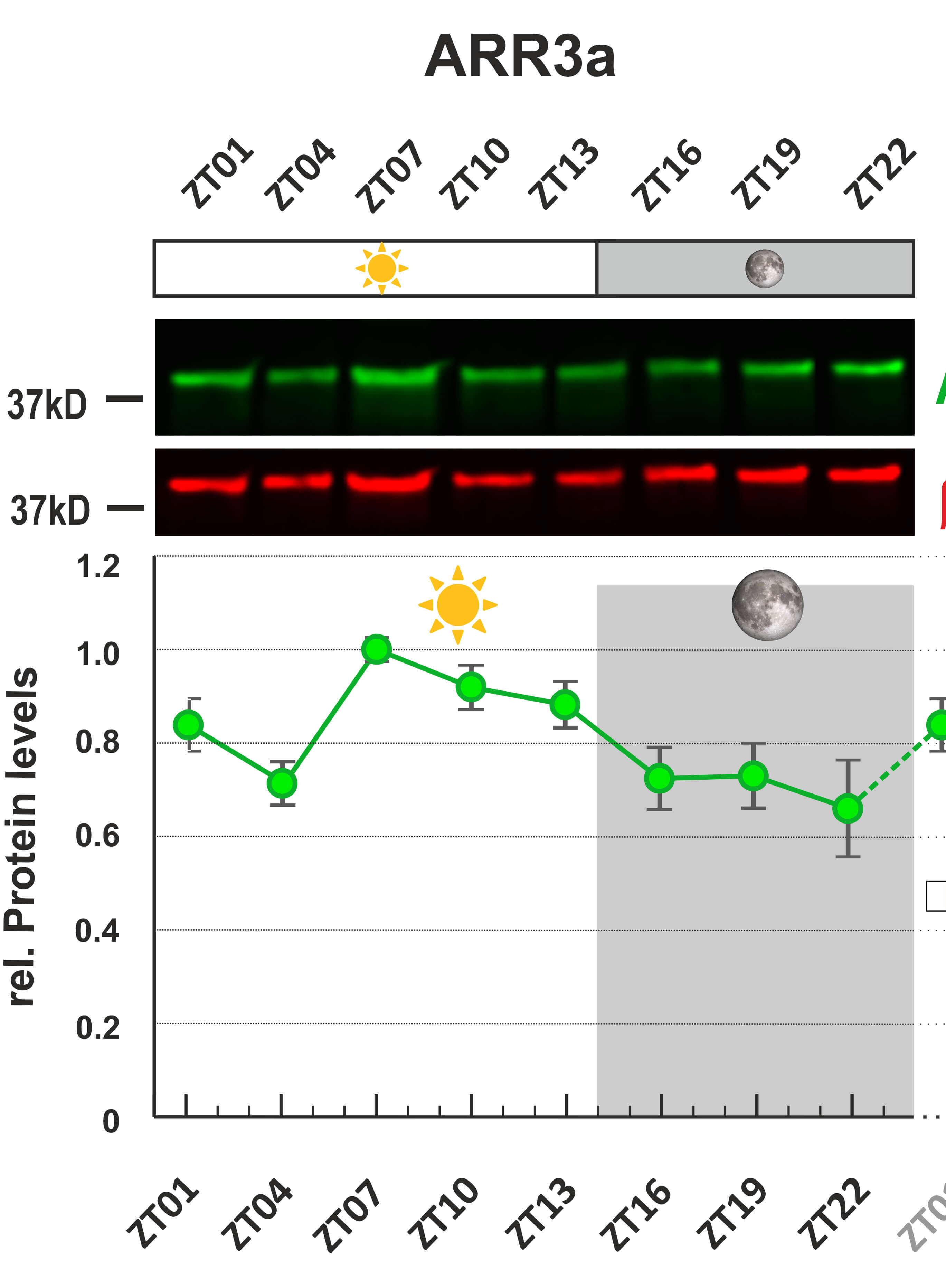
Detection of Zebrafish Retinal Proteins by Infrared Western Blotting

红外印迹法检测斑马鱼视网膜蛋白

JZ Jingjing Zang
SN Stephan Neuhauss
1339 Views
Feb 20, 2023
The zebrafish retina is a canonical vertebrate retina. Since the past few years, with the continually growing genetic toolbox and imaging techniques, zebrafish plays a crucial role in retinal research. This protocol describes a method to quantitatively evaluate the expression of Arrestin3a (Arr3a) and G-protein receptor kinase7a (Grk7a) in the adult zebrafish retina at protein levels by infrared fluorescence western blot. Our protocol can be easily adapted to measure protein levels in additional zebrafish tissues.

植物科学

水稻不同强度光刺激下叶绿体运动的成像

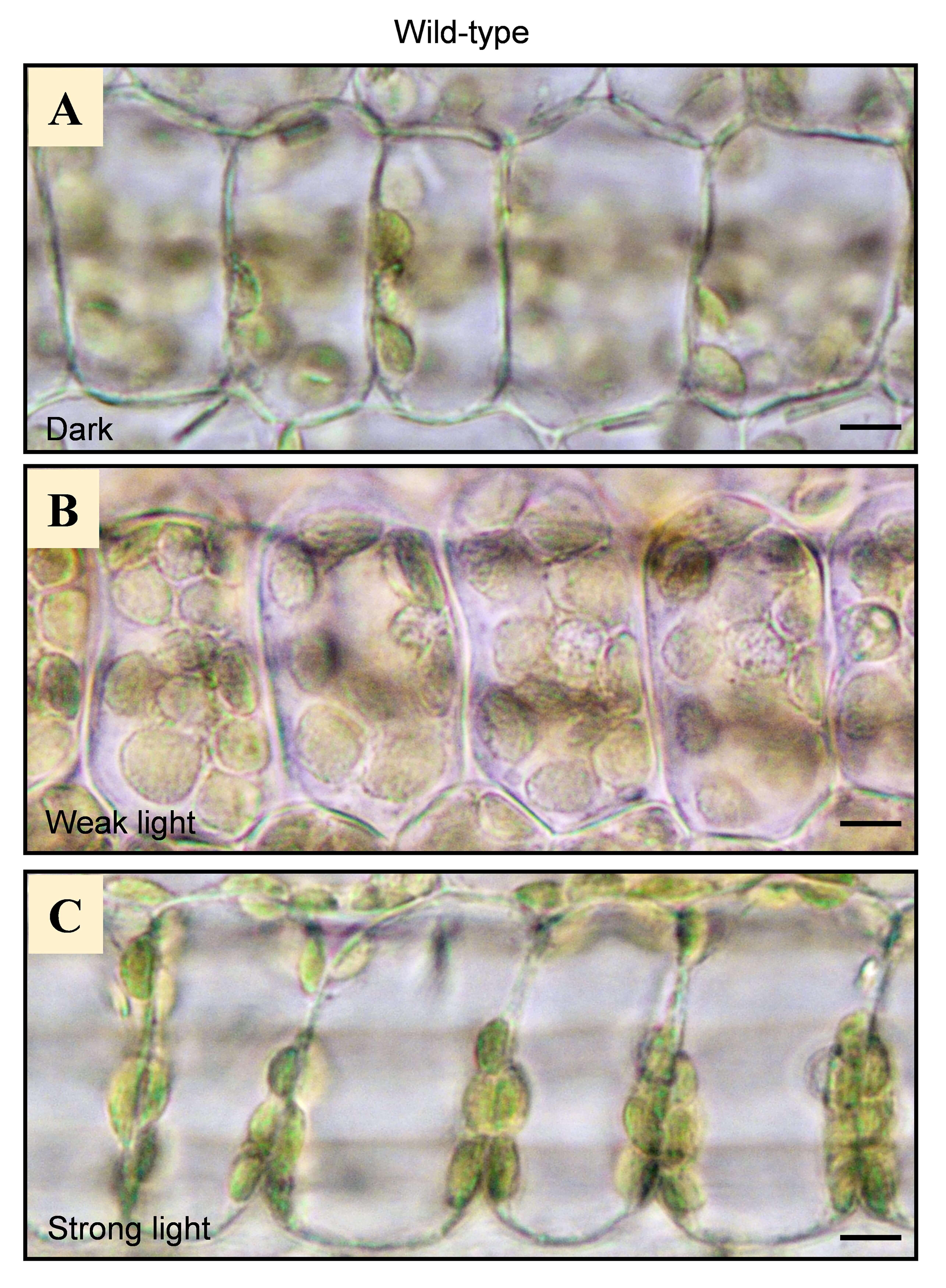
Imaging of Chloroplast Movement Responses to Light Stimulation in Different Intensities in Rice

水稻不同强度光刺激下叶绿体运动的成像

HW Hui Wu
ZW Ziyi Wang
YY Yanchun Yu
YZ Yanli Zhang
1014 Views
Feb 20, 2023
Chloroplast movement has been observed and analyzed since the 19th century. Subsequently, the phenomenon is widely observed in various plant species such as fern, moss, Marchantia polymorpha, and Arabidopsis. However, chloroplast movement in rice is less investigated, presumably due to the thick wax layer on its leaf surface, which reduces light sensitivity to the point that it was previously believed that there was no light-induced movement in rice. In this study, we present a convenient protocol suitable for observing chloroplast movement in rice only by optical microscopy without using special equipment. It will allow researchers to explore other signaling components involved in chloroplast movement in rice.