往期刊物2022

卷册: 12, 期号: 23

left

Dec

20

Dec

5

Nov

20

Nov

5

Oct

20

Oct

5

Sep

20

Sep

5

Aug

20

Aug

5

Jul

20

Jul

5

Jun

20

Jun

5

May

20

May

5

Apr

20

Apr

5

Mar

20

Mar

5

Feb

20

Feb

5

Jan

20

Jan

5

right

生物化学

利用 RNA Pull-down 技术评价蛋白质-RNA 相互作用的体外结合亲和力

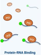
Assessing the in vitro Binding Affinity of Protein–RNA Interactions Using an RNA Pull-down Technique

利用 RNA Pull-down 技术评价蛋白质-RNA 相互作用的体外结合亲和力

AC Anand Chopra
FB Feras Balbous
KB Kyle K. Biggar
3215 Views
Dec 5, 2022
RNA is a vital component of the cell and is involved in a diverse range of cellular processes through a variety of functions. However, many of these functions cannot be performed without interactions with proteins. There are currently several techniques used to study protein–RNA interactions, such as electrophoretic mobility shift assay, fluorescence anisotropy, and filter binding. RNA-pulldown is a technique that uses biotinylated RNA probes to capture protein–RNA complexes of interest. First, the RNA probe and a recombinant protein are incubated to allow the in vitro interaction to occur. The fraction of bound protein is then captured by a biotin pull-down using streptavidin-agarose beads, followed by elution and immunoblotting for the recombinant protein with a His-tag–reactive probe. Overall, this method does not require specialized equipment outside what is typically found in a modern molecular laboratory and easily facilitates the maintenance of an RNase-free environment.Graphical abstract

癌症生物学

使用四甲基罗丹明甲酯染色对内吞死亡进行荧光延时成像

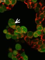
Fluorescence Time-lapse Imaging of Entosis Using Tetramethylrhodamine Methyl Ester Staining

使用四甲基罗丹明甲酯染色对内吞死亡进行荧光延时成像

EB Emir Bozkurt
HD Heiko Düssmann
JP Jochen H. M. Prehn
1208 Views
Dec 5, 2022
Entosis is a process where a living cell launches an invasion into another living cell’s cytoplasm. These inner cells can survive inside outer cells for a long period of time, can undergo cell division, or can be released. However, the fate of most inner cells is lysosomal degradation by entotic cell death. Entosis can be detected by imaging a combination of membrane, cytoplasmic, nuclear, and lysosomal staining in the cells. Here, we provide a protocol for detecting entosis events and measuring the kinetics of entotic cell death by time-lapse imaging using tetramethylrhodamine methyl ester (TMRM) staining.

免疫学

小鼠骨骼肌巨噬细胞富集的改善

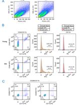
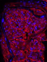
Improved Macrophage Enrichment from Mouse Skeletal Muscle

小鼠骨骼肌巨噬细胞富集的改善

LK Linda K. Krasniewski
DT Dimitrios Tsitsipatis
EI Elizabeth K. Izydore
CS Changyou Shi
YP Yulan Piao
MM Marc Michel
PS Payel Sen
MG Myriam Gorospe
CC Chang-Yi Cui
2161 Views
Dec 5, 2022
Macrophages are a heterogeneous class of innate immune cells that offer a primary line of defense to the body by phagocytizing pathogens, digesting them, and presenting the antigens to T and B cells to initiate adaptive immunity. Through specialized pro-inflammatory or anti-inflammatory activities, macrophages also directly contribute to the clearance of infections and the repair of tissue injury. Macrophages are distributed throughout the body and largely carry out tissue-specific functions. In skeletal muscle, macrophages regulate tissue repair and regeneration; however, the characteristics of these macrophages are not yet fully understood, and their involvement in skeletal muscle aging remains to be elucidated. To investigate these functions, it is critical to efficiently isolate macrophages from skeletal muscle with sufficient purity and yield for various downstream analyses. However, methods to prepare enriched skeletal muscle macrophages are scarce. Here, we describe in detail an optimized method to isolate skeletal muscle macrophages from mice. This method has allowed the isolation of CD45+/CD11b+ macrophage-enriched cells from young and old mice, which can be further used for flow cytometric analysis, fluorescence-activated cell sorting (FACS), and single-cell RNA sequencing.
用蛋白G亲和纯化法从大量血清样品中纯化人类自身 IgG

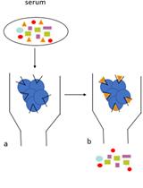
Human Auto-IgG Purification from High Volume Serum Sample by Protein G Affinity Purification

用蛋白G亲和纯化法从大量血清样品中纯化人类自身 IgG

SS Serena Sensi
AG Andreas Goebel
1977 Views
Dec 5, 2022
Immunoglobulins are proteins produced by the immune system, which bind specifically to the antigen that induced their formation and target it for destruction. Highly purified human immunoglobulins are commonly used in research laboratories for several applications, such as in vitro to obtain hybridomas and in vivo animal immunisation. Several affinity purification methods are used to purify immunoglobulins from human serum, such as protein A/G Sepharose beads, polyethylene glycol, and caprylic acid ammonium sulphate precipitation. Here, we provide a detailed protocol for purification of high-quality IgG from human serum, using affinity chromatography with protein G. The protocol is divided into four main steps (column preparation, serum running, wash, and elution) for IgG purification, and two extra steps (protein dialysis and sucrose concentration) that should be performed when buffer exchange and protein concentration are required. Several IgG affinity purification methods using protein A or G are available in the literature, but protein A has a higher affinity for rabbit, pig, dog, and cat IgG, while protein G has a higher affinity for mouse and human IgG. This affinity-based purification protocol uses protein G for a highly specific purification of human IgG for animal immunization, and it is particularly useful to purify large amounts of human IgG.Graphical abstractIgG purification protocol. The IgG purification protocol consists of four main steps (column preparation, serum running, wash, and elution) and two extra steps (protein dialysis and concentration). a. Diluted serum is added to the protein G beads and IgG binds to the Fc receptors on protein G beads. b. Beads are washed in Hartman’s solution to fully remove the complex protein mixture (multicolour shapes, as depicted in the graphical abstract). c. IgG (orange triangles, as depicted in the graphical abstract) are removed from protein G with glycine and collected in Tris buffer. d. The IgG is transferred into a semi-permeable membrane (‘snake skin’) and allowed to dialyse overnight for buffer exchange with a physiological solution (Hartmann’s).

医学

使用人外周血单核细胞模拟小鼠移植物对抗宿主疾病

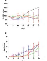
Modelling Graft-Versus-Host Disease in Mice Using Human Peripheral Blood Mononuclear Cells

使用人外周血单核细胞模拟小鼠移植物对抗宿主疾病

MH Manjurul Haque
DB Dominic A. Boardman
AL Avery J. Lam
KM Katherine N. MacDonald
LS Lieke Sanderink
QH Qing Huang
VF Vivian C. W. Fung
SI Sabine Ivison
MM Majid Mojibian
ML Megan K. Levings
2394 Views
Dec 5, 2022
Graft-versus-host disease (GvHD) is a significant complication of allogeneic hematopoietic stem cell transplantation. In order to develop new therapeutic approaches, there is a need to recapitulate GvHD effects in pre-clinical, in vivo systems, such as mouse and humanized mouse models. In humanized mouse models of GvHD, mice are reconstituted with human immune cells, which become activated by xenogeneic (xeno) stimuli, causing a multi-system disorder known as xenoGvHD. Testing the ability of new therapies to prevent or delay the development of xenoGvHD is often used as pre-clinical, proof-of-concept data, creating the need for standardized methodology to induce, monitor, and report xenoGvHD. Here, we describe detailed methods for how to induce xenoGvHD by injecting human peripheral blood mononuclear cells into immunodeficient NOD SCID gamma mice. We provide comprehensive details on methods for human T cell preparation and injection, mouse monitoring, data collection, interpretation, and reporting. Additionally, we provide an example of the potential utility of the xenoGvHD model to assess the biological activity of a regulatory T-cell therapy. Use of this protocol will allow better standardization of this model and comparison of datasets across different studies.Graphical abstract

分子生物学

通过斑点印迹法分析 N6-甲基腺苷 RNA 修饰水平

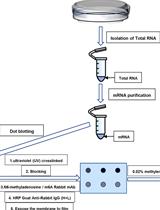
Analysis of N6-methyladenosine RNA Modification Levels by Dot Blotting

通过斑点印迹法分析 N6-甲基腺苷 RNA 修饰水平

YD Yu Du
MX Mingyue Xia
ZH Zhigang Hu
3415 Views
Dec 5, 2022
N6-methyladenosine (m6A) is the most prevalent internal modification of eukaryotic messenger RNAs (mRNAs), affecting their fold, stability, degradation, and cellular interaction(s) and implicating them in processes such as splicing, translation, export, and decay. The m6A modification is also extensively present in non-coding RNAs, including microRNAs (miRNAs), ribosomal RNAs (rRNAs), and transfer RNAs (tRNAs). Common m6A methylation detection techniques play an important role in understanding the biological function and potential mechanism of m6A, mainly including the quantification and specific localization of m6A modification sites. Here, we describe in detail the dot blotting method for detecting m6A levels in RNA (mRNA as an example), including total RNA extraction, mRNA purification, dot blotting, and data analysis. This protocol can also be used to enrich specific RNAs (such as tRNA, rRNA, or miRNA) by isolation technology to detect the m6A level of single RNA species, so as to facilitate further studies of the role of m6A in biological processes.

神经科学

哺乳动物真核和原核病原体对果蝇发育中枢神经系统的感染

Infection of the Developing Central Nervous System of Drosophila by Mammalian Eukaryotic and Prokaryotic Pathogens

哺乳动物真核和原核病原体对果蝇发育中枢神经系统的感染

BB Billel Benmimoun
BW Bente Winkler
PS Pauline Spéder
1326 Views
Dec 5, 2022
Pathogen invasion of the central nervous system (CNS) is an important cause of infection-related mortality worldwide and can lead to severe neurological sequelae. To gain access to the CNS cells, pathogens have to overcome the blood–brain barrier (BBB), a protective fence from blood-borne factors. To study host–pathogen interactions, a number of cell culture and animal models were developed. However, in vitro models do not recapitulate the 3D architecture of the BBB and CNS tissue, and in vivo mammalian models present cellular and technical complexities as well as ethical issues, rendering systematic and genetic approaches difficult. Here, we present a two-pronged methodology allowing and validating the use of Drosophila larvae as a model system to decipher the mechanisms of infection in a developing CNS. First, an ex vivo protocol based on whole CNS explants serves as a fast and versatile screening platform, permitting the investigation of molecular and cellular mechanisms contributing to the crossing of the BBB and consequences of infection on the CNS. Then, an in vivo CNS infection protocol through direct pathogen microinjection into the fly circulatory system evaluates the impact of systemic parameters, including the contribution of circulating immune cells to CNS infection, and assesses infection pathogenicity at the whole host level. These combined complementary approaches identify mechanisms of BBB crossing and responses of a diversity of CNS cells contributing to infection, as well as novel virulence factors of the pathogen.Graphical abstractProcedures flowchart. Mammalian neurotropic pathogens could be tested in two Drosophila central nervous system (CNS) infection setups (ex vivo and in vivo) for their ability to: (1) invade the CNS (pathogen quantifications), (2) disturb blood–brain barrier permeability, (3) affect CNS host cell behaviour (gene expression), and (4) alter host viability.
条件性舔抑制:评估独立于小鼠运动活动的情境、线索和情境线索复合恐惧反应

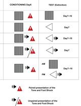
Conditioned Lick Suppression: Assessing Contextual, Cued, and Context-cue Compound Fear Responses Independently of Locomotor Activity in Mice

条件性舔抑制:评估独立于小鼠运动活动的情境、线索和情境线索复合恐惧反应

YB Youcef Bouchekioua
NN Naoya Nishitani
YO Yu Ohmura
1207 Views
Dec 5, 2022
Pavlovian fear conditioning is a widely used procedure to assess learning and memory processes that has also been extensively used as a model of post-traumatic stress disorder (PTSD). Freezing, the absence of movement except for respiratory-related movements, is commonly used as a measure of fear response in non-human animals. However, this measure of fear responses can be affected by a different baseline of locomotor activity between groups and/or conditions. Moreover, fear conditioning procedures are usually restricted to a single conditioned stimulus (e.g., a tone cue, the context, etc.) and thus do not depict the complexity of real-life situations where traumatic memories are composed of a complex set of stimuli associated with the same aversive event. To overcome this issue, we use a conditioned lick suppression paradigm where water-deprived mice are presented with a single conditioned stimulus (CS, a tone cue or the context) previously paired with an unconditioned stimulus (US, a foot shock) while consuming water. We use the ratio of number of licks before and during the CS presentation as a fear measure, thereby neutralizing the potential effect of locomotor activity in fear responses. We further implemented the conditioned lick suppression ratio to assess the effect of cue competition using a compound of contextual and tone cue conditioned stimuli that were extinguished separately. This paradigm should prove useful in assessing potential therapeutics and/or behavioral therapies in PTSD, while neutralizing potential confounding effects between locomotor activity and fear responses on one side, and by considering potential cue-competition effects on the other side.Graphical abstractSchematic representation of the compound context-cue condition lick suppression procedure. Illustration reproduced from Bouchekioua et al. (2022).

植物科学

用聚焦离子束铣削和低温电子断层扫描术方法研究洋葱原代细胞壁的结构

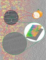
Focused Ion Beam Milling and Cryo-electron Tomography Methods to Study the Structure of the Primary Cell Wall in Allium cepa

用聚焦离子束铣削和低温电子断层扫描术方法研究洋葱原代细胞壁的结构

William J. Nicolas William J. Nicolas
GJ Grant J. Jensen
EM Elliot M. Meyerowitz
1761 Views
Dec 5, 2022
Cryo-electron tomography (cryo-ET) is a formidable technique to observe the inner workings of vitrified cells at a nanometric resolution in near-native conditions and in three-dimensions. One consequent drawback of this technique is the sample thickness, for two reasons: i) achieving proper vitrification of the sample gets increasingly difficult with sample thickness, and ii) cryo-ET relies on transmission electron microscopy (TEM), requiring thin samples for proper electron transmittance (<500 nm). For samples exceeding this thickness limit, thinning methods can be used to render the sample amenable for cryo-ET. Cryo-focused ion beam (cryo-FIB) milling is one of them and despite having hugely benefitted the fields of animal cell biology, virology, microbiology, and even crystallography, plant cells are still virtually unexplored by cryo-ET, in particular because they are generally orders of magnitude bigger than bacteria, viruses, or animal cells (at least 10 μm thick) and difficult to process by cryo-FIB milling. Here, we detail a preparation method where abaxial epidermal onion cell wall peels are separated from the epidermal cells and subsequently plunge frozen, cryo-FIB milled, and screened by cryo-ET in order to acquire high resolution tomographic data for analyzing the organization of the cell wall.
利用微滴数字 PCR 测量小麦植株中的转基因拷贝数

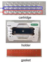
Measurement of Transgenes Copy Number in Wheat Plants Using Droplet Digital PCR

利用微滴数字 PCR 测量小麦植株中的转基因拷贝数

PL Peng Liu
SL Shuang Liu
JL Jiajia Lei
JC Jianping Chen
JY Jian Yang
1268 Views
Dec 5, 2022
Genetic transformation is a powerful method for the investigation of gene function and improvement of crop plants. The transgenes copy number in the transgenic line is involved in gene expression level and phenotypes. Additionally, identification of transgene zygosity is important for quantitative assessment of phenotype and for tracking the inheritance of transgenes in progeny generations. Several methods have been developed for estimating the transgene copy number, including southern blot assay and quantitative polymerase chain reaction (qPCR) experiments. Southern hybridization, although convincing and reliable, is a time-consuming method through which the examination of the copy number is challenging in species with large genomes like wheat plants. Although qPCR is potentially simpler to perform, its results lack accuracy and precision, especially to distinguish between one and two copy events in transgenic plants with large genomes. The droplet digital PCR (ddPCR)–based method for investigation of transgenes copy number has been widely used in an array of crops. In this method, the specific primers to amplify target transgenes and reference genes are used as a single duplexed reaction, which is divided into tens of thousands of nanodroplets. The copy number in independent transgenic lines is determined by detection and quantification of droplets using sequence-specific fluorescently labeled probes. This method offers superior accuracy and reliability with a low cost and scalability as other PCR techniques in the investigation of transgenes copy number.Graphical abstractFlow chart for the ddPCR protocol