往期刊物2022

卷册: 12, 期号: 22

left

Dec

20

Dec

5

Nov

20

Nov

5

Oct

20

Oct

5

Sep

20

Sep

5

Aug

20

Aug

5

Jul

20

Jul

5

Jun

20

Jun

5

May

20

May

5

Apr

20

Apr

5

Mar

20

Mar

5

Feb

20

Feb

5

Jan

20

Jan

5

right

生物工程

一种快速、高效的组织切片去细胞方法

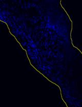
A Fast and Efficient Decellularization Method for Tissue Slices

一种快速、高效的组织切片去细胞方法

MN Maria Narciso
AU Anna Ulldemolins
CJ Constança Júnior
JO Jorge Otero
DN Daniel Navajas
RF Ramon Farré
NG Núria Gavara
IA Isaac Almendros
2146 Views
Nov 20, 2022
The study and use of decellularized extracellular matrix (dECM) in tissue engineering, regenerative medicine, and pathophysiology have become more prevalent in recent years. To obtain dECM, numerous decellularization procedures have been developed for the entire organ or tissue blocks, employing either perfusion of decellularizing agents through the tissue’s vessels or submersion of large sections in decellularizing solutions. However, none of these protocols are suitable for thin tissue slices (less than 100 µm) or allow side-by-side analysis of native and dECM consecutive tissue slices. Here, we present a detailed protocol to decellularize tissue sections while maintaining the sample attached to a glass slide. This protocol consists of consecutive washes and incubations of simple decellularizing agents: ultrapure water, sodium deoxycholate (SD) 2%, and deoxyribonuclease I solution 0.3 mg/mL (DNase I). This novel method has been optimized for a faster decellularization time (2–3 h) and a better correlation between dECM properties and native tissue-specific biomarkers, and has been tested in different types of tissues and species, obtaining similar results. Furthermore, this method can be used for scarce and valuable samples such as clinical biopsies.

发育生物学

监测秀丽隐杆线虫凋亡细胞清除过程中自噬体与吞噬体的募集与融合

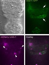
Monitoring the Recruitment and Fusion of Autophagosomes to Phagosomes During the Clearance of Apoptotic Cells in the Nematode Caenorhabditis elegans

监测秀丽隐杆线虫凋亡细胞清除过程中自噬体与吞噬体的募集与融合

OP Omar Peña-Ramos
ZZ Zheng Zhou
1507 Views
Nov 20, 2022
During an animal's development, a large number of cells undergo apoptosis, a suicidal form of death. These cells are promptly phagocytosed by other cells and degraded inside phagosomes. The recognition, engulfment, and degradation of apoptotic cells is an evolutionarily conserved process occurring in all metazoans. Recently, we discovered a novel event in the nematode Caenorhabditis elegans: the double-membrane autophagosomes are recruited to the surface of phagosomes; subsequently, the outer membrane of an autophagosome fuses with the phagosomal membrane, allowing the inner vesicle to enter the phagosomal lumen and accumulate there over time. This event facilitates the degradation of the apoptotic cell inside the phagosome. During this study, we developed a real-time imaging protocol monitoring the recruitment and fusion of autophagosomes to phagosomes over two hours during embryonic development. This protocol uses a deconvolution-based microscopic imaging system with an optimized setting to minimize photodamage of the embryo during the recording period for high-resolution images. Furthermore, acid-resistant fluorescent reporters are chosen to label autophagosomes, allowing the inner vesicles of an autophagosome to remain visible after entering the acidic phagosomal lumen. The methods described here, which enable high sensitivity, quantitative measurement of each step of the dynamic incorporation in developing embryos, are novel since the incorporation of autophagosomes to phagosomes has not been reported previously. In addition to studying the degradation of apoptotic cells, this protocol can be applied to study the degradation of non-apoptotic cell cargos inside phagosomes, as well as the fusion between other types of intracellular organelles in living C. elegans embryos. Furthermore, its principle of detecting the membrane fusion event can be adapted to study the relationship between autophagosomes and phagosomes or other intracellular organelles in any biological system in which real-time imaging can be conducted.

药物发现

评估离子载体阳离子转运的体外检测方法

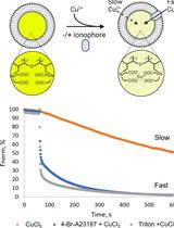
In vitro Assay to Evaluate Cation Transport of Ionophores

评估离子载体阳离子转运的体外检测方法

HU Huriye D. Uzun
MV Melissa Vázquez-Hernández
JB Julia E. Bandow
TP Thomas Günther Pomorski
1655 Views
Nov 20, 2022
Ion homeostasis is a fundamental regulator of cellular processes and depends upon lipid membranes, which function as ion permeability barriers. Ionophores facilitate ion transport across cell membranes and offer a way to manipulate cellular ion composition. Here, we describe a calcein quenching assay based on large unilamellar vesicles that we used to evaluate divalent cation transport of the ionophore 4-Br-A23187. This assay can be used to study metal transport by ionophores and membrane proteins, under well-defined conditions.Graphical abstract:

微生物学

用于检测和量化生化物质、代谢物和天然产物的荧光结合蛋白传感器

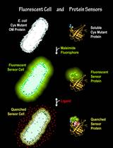
Fluorescent Binding Protein Sensors for Detection and Quantification of Biochemicals, Metabolites, and Natural Products

用于检测和量化生化物质、代谢物和天然产物的荧光结合蛋白传感器

SN Salete M. Newton
PK Phillip E. Klebba
2099 Views
Nov 20, 2022
Membrane transporters and soluble binding proteins recognize particular nutrients, metabolites, vitamins, or ligands. By modifying genetically engineered single cysteine residues near the active sites of such proteins with extrinsic maleimide fluorophores, the approaches that we report create sensitive fluorescent sensors that detect, quantify, and monitor molecules that are relevant to the biochemistry, physiology, microbiology, and clinical properties of pro- and eukaryotic organisms.Graphical abstract:
促进巴贝虫生物学、发病机制和治疗的巴贝虫培养和小鼠(ICIM)模型

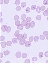
Babesia duncani in Culture and in Mouse (ICIM) Model for the Advancement of Babesia Biology, Pathogenesis and Therapy

促进巴贝虫生物学、发病机制和治疗的巴贝虫培养和小鼠(ICIM)模型

VK Vandana Kumari
AP Anasuya C. Pal
PS Pallavi Singh
CB Choukri Ben Mamoun
1318 Views
Nov 20, 2022
Babesiosis is a tick-borne disease caused by pathogens belonging to the genus Babesia. In humans, the disease presents as a malaria-like illness and can be fatal in immunocompromised and elderly people. In the past few years, human babesiosis has been a rising concern worldwide. The disease is transmitted through tick bite, blood transfusion, and transplacentally in rare cases, with several species of Babesia causing human infection. Babesia microti, Babesia duncani, and Babesia divergens are of particular interest because of their important health impact and amenability to research inquiries. B. microti, the most commonly reported Babesia pathogen infecting humans, can be propagated in immunocompetent and immunocompromised mice but so far has not been successfully continuously propagated in vitro in human red blood cells (hRBCs). Conversely, B. divergens can be propagated in vitro in hRBCs but lacks a mouse model to study its virulence. Recent studies have highlighted the uniqueness of B. duncani as an ideal model organism to study intraerythrocytic parasitism in vitro and in vivo. An optimized B. duncani in culture and in mouse (ICIM) model has recently been described, combining long-term continuous in vitro culture of the parasite in human red blood cells with an animal model of parasitemia (P) and lethal infection in C3H/HeJ mice. Here, we provide a detailed protocol for the use of the B. duncani ICIM model in research. This model provides a unique and sound foundation to gain further insights into the biology, pathogenesis, and virulence of Babesia and other intraerythrocytic parasites, and has been validated as an efficient system to evaluate novel strategies for the treatment of human babesiosis and possibly other parasitic diseases.Graphical abstract: ICIM model [Adapted and modified from Pal et al. (2022)]
利用改进的培养方法和生长监测对嗜盐火山生菌进行表型分析的次氯酸盐胁迫试验

Hypochlorite Stress Assay for Phenotypic Analysis of the Halophilic Archaeon Haloferax volcanii Using an Improved Incubation Method and Growth Monitoring

利用改进的培养方法和生长监测对嗜盐火山生菌进行表型分析的次氯酸盐胁迫试验

PM Paula Mondragon
SH Sungmin Hwang
AS Amy Schmid
JM Julie A. Maupin-Furlow
1079 Views
Nov 20, 2022
The study of haloarchaea provides an opportunity to expand understanding of the mechanisms used by extremophiles to thrive in and respond to harsh environments, including hypersaline and oxidative stress conditions. A common strategy used to investigate molecular mechanisms of stress response involves the deletion and/or site-directed mutagenesis of genes identified through omics studies followed by a comparison of the mutant and wild-type strains for phenotypic differences. The experimental methods used to monitor these differences must be controlled and reproducible. Current methods to examine recovery of halophilic archaea from extreme stress are complicated by extended incubation times, nutrients not typically encountered in the environment, and other related limitations. Here we describe a method for assessing the function of genes during hypochlorite stress in the halophilic archaeon Haloferax volcanii that overcomes these types of limitations. The method was found reproducible and informative in identifying genes needed for H. volcanii to recover from hypochlorite stress.

神经科学

用于识别亚细胞和超细胞药代动力学的中枢神经系统作用的药物-生物传感器配对的荧光筛选

Fluorescence Screens for Identifying Central Nervous System–Acting Drug–Biosensor Pairs for Subcellular and Supracellular Pharmacokinetics

用于识别亚细胞和超细胞药代动力学的中枢神经系统作用的药物-生物传感器配对的荧光筛选

ZB Zoe G. Beatty
AM Anand K. Muthusamy
EU Elizabeth K. Unger
DD Dennis A. Dougherty
LT Lin Tian
LL Loren L. Looger
AS Amol V. Shivange
KB Kallol Bera
HL Henry A. Lester
AN Aaron L. Nichols
2109 Views
Nov 20, 2022
Subcellular pharmacokinetic measurements have informed the study of central nervous system (CNS)–acting drug mechanisms. Recent investigations have been enhanced by the use of genetically encoded fluorescent biosensors for drugs of interest at the plasma membrane and in organelles. We describe screening and validation protocols for identifying hit pairs comprising a drug and biosensor, with each screen including 13–18 candidate biosensors and 44–84 candidate drugs. After a favorable hit pair is identified and validated via these protocols, the biosensor is then optimized, as described in other papers, for sensitivity and selectivity to the drug. We also show sample hit pair data that may lead to future intensity-based drug-sensing fluorescent reporters (iDrugSnFRs). These protocols will assist scientists to use fluorescence responses as criteria in identifying favorable fluorescent biosensor variants for CNS-acting drugs that presently have no corresponding biosensor partner. Graphical abstract:
用于动态和电泳光散射测量的均质肌动蛋白丝的体外制备

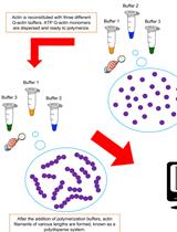
In vitro Preparation of Homogenous Actin Filaments for Dynamic and Electrophoretic Light Scattering Measurements

用于动态和电泳光散射测量的均质肌动蛋白丝的体外制备

EA Ernesto Alva
AG Annitta George
LB Lorenzo Brancaleon
MM Marcelo Marucho
1174 Views
Nov 20, 2022
Actin filaments are essential for various biological activities in eukaryotic cellular processes. Available in vitro experimental data on these systems often lack details and information on sample preparation protocols and experimental techniques, leading to unreproducible results. Additionally, different experimental techniques and polymerization buffers provide different, sometimes contradictory results on the properties of these systems, making it substantially difficult to gather meaningful data and conclusive information from them. This article presents a robust, accurate, detailed polymerization protocol to prepare high-quality actin filament samples for light scattering experiments. It has been shown to provide unicity and consistency in preparing stable, dispersed, aggregates-free, homogenous actin filament samples that could benefit many other scientific research groups currently working in the field. To develop the protocol, we used conventional actin buffers in physiological conditions. However, it can easily be adapted to prepare samples using other buffers and biological fluids. This protocol yielded reproducible results on essential actin filament parameters such as the translational diffusion coefficient and electrophoretic mobility. Overall, suitable modifications of the proposed experimental method could generate accurate, reproducible light scattering results on other highly charged anionic filaments commonly found in biological cells (e.g., microtubules, DNAs, RNAs, or filamentous viruses).Graphical abstract:

干细胞

使用BODIPY FL-鞘脂类化合物研究小鼠胚胎干细胞中的鞘脂类代谢

Using BODIPY FL-Sphingolipid Analogs to Study Sphingolipid Metabolism in Mouse Embryonic Stem Cells

使用BODIPY FL-鞘脂类化合物研究小鼠胚胎干细胞中的鞘脂类代谢

WF Wei Fan
XL Xiaoling Li
1750 Views
Nov 20, 2022
Sphingolipids are important structural components of cellular membranes. They also function as prominent signaling molecules to control a variety of cellular events, such as cell growth, differentiation, and apoptosis. Impaired sphingolipid metabolism, particularly defects in sphingolipid degradation, has been associated with many human diseases. Fluorescence sphingolipid analogs have been widely used as efficient probes to study sphingolipid metabolism and intracellular trafficking in living mammalian cells. Compared with nitrobenzoxadiazole fluorophores (NBD FL), the boron dipyrromethene difluoride fluorophores (BODIPY FL) have much higher absorptivity and fluorescence quantum. These features allow more intensive labeling of cells for fluorescence microscopy imaging and flow cytometry analysis. Here, we describe a protocol employing BODIPY FL-labeled sphingolipid analogs to elucidate sphingolipid internalization, trafficking, and endocytosis in mouse embryonic stem cells.Graphical abstract:

系统生物学

在哺乳动物细胞中进行蛋白质组积分溶解度改变(PISA)测定,用于深度、高置信度和高通量目标解构

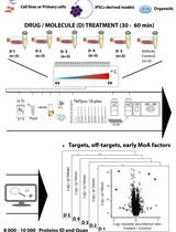
Proteome Integral Solubility Alteration (PISA) Assay in Mammalian Cells for Deep, High-Confidence, and High-Throughput Target Deconvolution

在哺乳动物细胞中进行蛋白质组积分溶解度改变(PISA)测定,用于深度、高置信度和高通量目标解构

XZ Xuepei Zhang
OL Olga Lytovchenko
SL Susanna L. Lundström
RZ Roman A. Zubarev
MG Massimiliano Gaetani
3508 Views
Nov 20, 2022
Chemical proteomics focuses on the drug–target–phenotype relationship for target deconvolution and elucidation of the mechanism of action—key and bottleneck in drug development and repurposing. Majorly due to the limits of using chemically modified ligands in affinity-based methods, new, unbiased, proteome-wide, and MS-based chemical proteomics approaches have been developed to perform drug target deconvolution, using full proteome profiling and no chemical modification of the studied ligand. Of note among them, thermal proteome profiling (TPP) aims to identify the target(s) by measuring the difference in melting temperatures between each identified protein in drug-treated versus vehicle-treated samples, with the thermodynamic interpretation of “protein melting” and curve fitting of all quantified proteins, at all temperatures, in each biological replicate. Including TPP, all the other chemical proteomics approaches often fail to provide target deconvolution with sufficient proteome depth, statistical power, throughput, and sustainability, which could hardly fulfill the final purpose of drug development. The proteome integral solubility alteration (PISA) assay provides no thermodynamic interpretation, but a throughput 10–100-fold compared to the other proteomics methods, high sustainability, much lower time of analysis and sample amount requirements, high confidence in results, maximal proteome coverage (~10,000 protein IDs), and up to five drugs / test molecules in one assay, with at least biological triplicates of each treatment. Each drug-treated or vehicle-treated sample is split into many fractions and exposed to a gradient of heat as solubility perturbing agent before being recomposed into one sample; each soluble fraction is isolated, then deep and quantitative proteomics is applied across all samples. The proteins interacting with the tested molecules (targets and off-targets), the activated mechanistic factors, or proteins modified during the treatment show reproducible changes in their soluble amount compared to vehicle-treated controls. As of today, the maximal multiplexing capability is 18 biological samples per PISA assay, which enables statistical robustness and flexible experimental design accommodation for fuller target deconvolution, including integration of orthogonal chemical proteomics methods in one PISA assay. Living cells for studying target engagement in vivo or, alternatively, protein extracts to identify in vitro ligand-interacting proteins can be studied, and the minimal need in sample amount unlocks target deconvolution using primary cells and their derived cultures.Graphical abstract:
利用Gibson组装技术高效生成蛋白质配体筛选全基因组文库

Efficient Generation of Genome-wide Libraries for Protein–ligand Screens Using Gibson Assembly

利用Gibson组装技术高效生成蛋白质配体筛选全基因组文库

TS Tamara Sternlieb
ML Mira Loock
MG Mengjin Gao
IC Igor Cestari
2491 Views
Nov 20, 2022
Genome-wide screens using yeast or phage displays are powerful tools for identifying protein–ligand interactions, including drug or vaccine targets, ligand receptors, or protein–protein interactions. However, assembling libraries for genome-wide screens can be challenging and often requires unbiased cloning of 105–107 DNA fragments for a complete representation of a eukaryote genome. A sub-optimal genomic library can miss key genomic sequences and thus result in biased screens. Here, we describe an efficient method to generate genome-wide libraries for yeast surface display using Gibson assembly. The protocol entails genome fragmentation, ligation of adapters, library cloning using Gibson assembly, library transformation, library DNA recovery, and a streamlined Oxford nanopore library sequencing procedure that covers the length of the cloned DNA fragments. We also describe a computational pipeline to analyze the library coverage of the genome and predict the proportion of expressed proteins. The method allows seamless library transfer among multiple vectors and can be easily adapted to any expression system.