往期刊物2022

卷册: 12, 期号: 19

left

Dec

20

Dec

5

Nov

20

Nov

5

Oct

20

Oct

5

Sep

20

Sep

5

Aug

20

Aug

5

Jul

20

Jul

5

Jun

20

Jun

5

May

20

May

5

Apr

20

Apr

5

Mar

20

Mar

5

Feb

20

Feb

5

Jan

20

Jan

5

right

生物工程

双特异性抗体双靶点桥接ELISA

Dual-target Bridging ELISA for Bispecific Antibodies

双特异性抗体双靶点桥接ELISA

MP Min Pei
YW Yao Wang
LT Lei Tang
WW Weitao Wu
CW Chunhe Wang
YC Yi-Li Chen
3328 Views
Oct 5, 2022
Bispecific antibodies (BsAbs) are typically monoclonal antibody (mAb)–derived molecular entities engineered to bind to two distinct targets, including two antigens or two epitopes on the same antigen. When compared to parental monoclonal antibodies or combinational therapies, the generated BsAbs have the ability to bridge the two targets and thus may offer additional clinical benefits. Characterizing BsAbs’ ability to bind to both targets simultaneously is critical for their biotherapeutic development. A range of bi-functional quantitative bridging assays to enable target-specific capture and detection of binding properties include enzyme-linked immunosorbent assay (ELISA), surface plasmon resonance (SPR), and cell-based flow cytometry. Developing suitable and robust cell-based bioassays is more challenging than non-cell-based binding assays because cell-based assays with complex matrices can be inherently variable and often lack precision. Compared to SPR, ELISA has a rapid setup and readily available method, being widely and extensively applied in almost every laboratory. Here, we describe a dual-target bridging ELISA assay that characterizes the ability of a HER2(human epidermal growth factor receptor 2)/PD-L1(programmed cell death ligand 1) BsAb in binding to both HER2 and PD-L1 simultaneously, a prerequisite for its envisioned mode of action.Graphical abstract:
LIST:新开发的激光辅助细胞生物打印技术

LIST: A Newly Developed Laser-assisted Cell Bioprinting Technology

LIST:新开发的激光辅助细胞生物打印技术

KR Katiane Roversi
HE Hamid Ebrahimi Orimi
ME Mahyar Erfanian
ST Sebastien Talbot
CB Christos Boutopoulos
1340 Views
Oct 5, 2022
Cell bioprinting technologies aim to fabricate tissue-like constructs by delivering biomaterials layer-by-layer. Bioprinted constructs can reduce the use of animals in drug development and hold promise for addressing the shortage of organs for transplants. We recently introduced a laser-assisted drop-on-demand bioprinting technology termed Laser Induced Side Transfer (LIST). This technology can print delicate cell types, including primary neurons. This bioprinting protocol includes the following key steps: cell harvesting, bio-ink preparation, laser setup priming, printing, and post-printing analysis. This protocol includes a detailed description of the laser setup, which is a rather unusual setup for a biology lab. This should allow easy reproduction by readers with basic knowledge of optics. Although we have focused on neuron bioprinting, interested readers will be able to adapt the protocol to bioprint virtually any cell type.Graphical abstract:

细胞生物学

HuTu-80 和 HEK 293T 细胞系中 CRISPR/Cas9 介导的 LRP10 敲除

CRISPR/Cas9-mediated LRP10 Knockout in HuTu-80 and HEK 293T Cell Lines

HuTu-80 和 HEK 293T 细胞系中 CRISPR/Cas9 介导的 LRP10 敲除

MG Martyna M. Grochowska
VB Vincenzo Bonifati
WM Wim Mandemakers
2898 Views
Oct 5, 2022
Loss-of-function (LoF) variants in the low-density lipoprotein receptor–related protein 10 gene (LRP10) have been recently implicated in the development of neurodegenerative diseases, including Parkinson's disease (PD), PD dementia (PDD), and dementia with Lewy bodies (DLB). However, despite the genetic evidence, little is known about the LRP10 protein function in health and disease. Here, we describe a detailed protocol to efficiently generate a LRP10 LoF model in two independent LRP10-expressing cell lines, HuTu-80 and HEK 293T, using the CRISPR/Cas9 genome-editing tool. Our method efficiently generates bi-allelic LRP10 knockout (KO), which can be further utilized to elucidate the physiological LRP10 protein function and to model some aspects of neurodegenerative disorders. Graphical abstract: CRISPR/Cas9 workflow for the generation of the LRP10 KO. (1) Designed single guide RNA (sgRNA) is cloned into CRISPR/Cas9 px458 plasmid. (2) Cells are transfected with the CRISPR/Cas9 plasmid containing sgRNA. (3) Two days post transfection, cells are dissociated and sorted as single cells by fluorescence-activated cell sorting (FACS). (4) After several weeks, expanded clonal lines are (5) verified with Sanger sequencing for the presence of INDELs (insertions or deletions), RT-qPCR for the amounts of LRP10 mRNA transcript, and Western blotting for the analysis of the LRP10 protein levels.
用于鉴定核糖核蛋白复合物中 mRNA 种类的增强型核糖核蛋白免疫沉淀 (RIP) 技术

Enhanced Ribonucleoprotein Immunoprecipitation (RIP) Technique for the Identification of mRNA Species in Ribonucleoprotein Complexes

用于鉴定核糖核蛋白复合物中 mRNA 种类的增强型核糖核蛋白免疫沉淀 (RIP) 技术

SF Saja A. Fakhraldeen
SB Scott M. Berry
DB David J. Beebe
CA Caroline M. Alexander
1959 Views
Oct 5, 2022
RNA binding proteins (RBPs) are critical regulators of cellular phenotypes, and dysregulated RBP expression is implicated in various diseases including cancer. A single RBP can bind to and regulate the expression of many RNA molecules via a variety of mechanisms, including translational suppression, prevention of RNA degradation, and alteration in subcellular localization. To elucidate the role of a specific RBP within a given cellular context, it is essential to first identify the group of RNA molecules to which it binds. This has traditionally been achieved using cross-linking-based assays in which cells are first exposed to agents that cross-link RBPs to nucleic acids and then lysed to extract and purify the RBP-nucleic acid complexes. The nucleic acids within the mixture are then released and analyzed via conventional means (e.g., microarray analysis, qRT-PCR, RNA sequencing, or Northern blot). While cross-linking-based ribonucleoprotein immunoprecipitation (RIP) has proven its utility within some contexts, it is technically challenging, inefficient, and suboptimal given the amount of time and resources (e.g., cells and antibodies) required. Additionally, these types of studies often require the use of over-expressed versions of proteins, which can introduce artifacts. Here, we describe a streamlined version of RIP that utilizes exclusion-based purification technologies. This approach requires significantly less starting material and resources compared to traditional RIP approaches, takes less time, which is tantamount given the labile nature of RNA, and can be used with endogenously expressed proteins. The method described here can be used to study RNA-protein interactions in a variety of cellular contexts.Graphical abstract:

药物发现

原肠胚筛选以识别斑马鱼胚胎中的抗转移药物

Gastrulation Screening to Identify Anti-metastasis Drugs in Zebrafish Embryos

原肠胚筛选以识别斑马鱼胚胎中的抗转移药物

JN Joji Nakayama
HM Hideki Makinoshima
ZG Zhiyuan Gong
1705 Views
Oct 5, 2022
Few models exist that allow for rapid and effective screening of anti-metastasis drugs. Here, we present a drug screening protocol utilizing gastrulation of zebrafish embryos for identification of anti-metastasis drugs. Based on the evidence that metastasis proceeds through utilizing the molecular mechanisms of gastrulation, we hypothesized that chemicals interrupting zebrafish gastrulation might suppress the metastasis of cancer cells. Thus, we developed a phenotype-based chemical screen that uses epiboly, the first morphogenetic movement in gastrulation, as a marker. The screen only needs zebrafish embryos and enables hundreds of chemicals to be tested in five hours by observing the epiboly progression of chemical-treated embryos. In the screen, embryos at the two-cell stage are firstly corrected and then developed to the sphere stage. The embryos are treated with a test chemical and incubated in the presence of the chemical until vehicle-treated embryos develop to the 90% epiboly stage. Finally, positive ‘hit’ chemicals that interrupt epiboly progression are selected by comparing epiboly progression of the chemical-treated and vehicle-treated embryos under a stereoscopic microscope. A previous study subjected 1,280 FDA-approved drugs to the screen and identified adrenosterone and pizotifen as epiboly-interrupting drugs. These were validated to suppress metastasis of breast cancer cells in mice models of metastasis. Furthermore, 11β-hydroxysteroid dehydrogenase 1 (HSD11β1) and serotonin receptor 2C (HTR2C), the primary targets of adrenosterone and pizotifen, respectively, promoted metastasis through induction of epithelial-mesenchymal transition (EMT). Therefore, this screen could be converted into a chemical genetic screening platform for identification of metastasis-promoting genes.Graphical abstract:

免疫学

腹腔巨噬细胞中 SIRT6 表达的流式细胞术分析

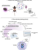
Flow Cytometry Analysis of SIRT6 Expression in Peritoneal Macrophages

腹腔巨噬细胞中 SIRT6 表达的流式细胞术分析

VP Valentina Pérez-Torrado
JR Jorge Rodríguez-Duarte
EC Carlos Escande
MB Mariana Bresque
2833 Views
Oct 5, 2022
The sirtuin 6 has emerged as a regulator of acute and chronic immune responses. Recent findings show that SIRT6 is necessary for mounting an active inflammatory response in macrophages. In vitro studies revealed that SIRT6 is stabilized in the cytoplasm to promote tumor necrosis factor (TNFα) secretion. Notably, SIRT6 also promotes TNFα secretion by resident peritoneal macrophages upon lipopolysaccharide (LPS) stimulation in vivo. Although many studies have investigated SIRT6 function in the immune response through different genetic and pharmacological approaches, direct measurements of in vivo SIRT6 expression in immune cells by flow cytometry have not yet been performed. Here, we describe a step-by-step protocol for peritoneal fluid extraction, isolation, and preparation of peritoneal cavity cells, intracellular SIRT6 staining, and flow cytometry analysis to measure SIRT6 levels in mice peritoneal macrophages. By providing a robust method to quantify SIRT6 levels in different populations of macrophages, this method will contribute to deepening our understanding of the role of SIRT6 in immunity, as well as in other cellular processes regulated by SIRT6.Graphical abstract:

微生物学

蛋白酶样蛋白酶抑制剂活性测定

Assay for Protealysin-like Protease Inhibitor Activity

蛋白酶样蛋白酶抑制剂活性测定

IB Igor M. Berdyshev
MK Maria A. Karaseva
ID Ilya V. Demidyuk
1461 Views
Oct 5, 2022
Here, we present the first quantitative method for the activity analysis of protealysin-like protease (PLP) inhibitors. This approach is based on a previously developed method for protealysin activity determination by hydrolysis of internally quenched fluorescent peptide substrate 2-aminobenzoyl-L-arginyl-L-seryl-L-valyl-L-isoleucyl-L-(ϵ-2,4-dinitrophenyl)lysine. In this protocol, we significantly reduced enzyme concentration and introduced some minor modifications to decrease variation between replicates. The protocol was validated using emfourin, a novel proteinaceous metalloprotease inhibitor. Data obtained demonstrates that the developed assay method is an affordable approach for characterizing and screening various PLP inhibitors.Graphical abstract:

神经科学

妊娠晚期短暂性产前缺氧的简化范式研究发育性低氧损伤的功能和结构结果

A Simplified Paradigm of Late Gestation Transient Prenatal Hypoxia to Investigate Functional and Structural Outcomes from a Developmental Hypoxic Insult

妊娠晚期短暂性产前缺氧的简化范式研究发育性低氧损伤的功能和结构结果

EG Elyse C. Gadra
AC Ana G. Cristancho
1706 Views
Oct 5, 2022
Late-gestation transient intrauterine hypoxia is a common cause of birth injury. It can lead to long-term neurodevelopmental disabilities even in the absence of gross anatomic injury. Currently, postnatal models of hypoxia–ischemia are most commonly used to study the effect of oxygen deprivation in the fetal brain. These models, however, are unable to take into account placental factors that influence the response to hypoxia, exhibit levels of cell death not seen in many human patients, and are unable to model preterm hypoxia. To address this gap in research, we have developed a protocol to induce transient hypoxia in fetal mice. A pregnant dam at gestational day 17.5 is placed into a hypoxia chamber. Over 30 min, the inspired oxygen is titrated from 21% (ambient air) to 5%. The dam remains in the chamber for up to 8 h, after which fetal brains can be collected or pups delivered for postnatal studies. This protocol recapitulates phenotypes seen in human patients exposed to transient in utero hypoxia and is readily reproducible by researchers.Graphical abstract:

植物科学

豌豆腋芽植物激素和 RNA 的提取与定量

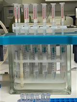
Extraction and Quantification of Plant Hormones and RNA from Pea Axillary Buds

豌豆腋芽植物激素和 RNA 的提取与定量

DC Da Cao
FB Francois Barbier
KY Kaori Yoneyama
CB Christine A. Beveridge
2198 Views
Oct 5, 2022
The quantification of plant hormones and related gene expression is essential to improve the understanding of the molecular regulation of plant growth and development. However, plant hormone quantification is still challenging due to extremely low endogenous levels and high chemical diversity. In this study, we present a convenient extraction protocol that enables the simultaneous extraction of both phytohormones and RNA from the same sample in a small quantity (approximately 10 mg). Using ultra-performance liquid chromatography coupled with tandem mass spectrometry (UPLC–MS/MS), this protocol provides a method to quantify 13 phytohormones and their derivatives from four classes (cytokinin, auxin, abscisic acid, and gibberellin) at the speed of 14 min per sample.
从模式植物拟南芥和农作物大豆中收集木质部分泌物

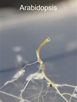
Collection of Xylem Exudates from the Model Plant Arabidopsis and the Crop Plant Soybean

从模式植物拟南芥和农作物大豆中收集木质部分泌物

SO Satoru Okamoto
AK Azusa Kawasaki
2101 Views
Oct 5, 2022
A number of molecules, such as secreted peptides, have been shown to mediate root-to-shoot signaling in response to various conditions. The xylem is a pathway for water and molecules that are translocated from roots to shoots. Therefore, collecting and analyzing xylem exudates is an efficient approach to study root-to-shoot long-distance signaling. Here, we describe a step-by-step protocol for the collection of xylem exudate from the model plant Arabidopsis and the crop plant soybean (Glycine max). In this protocol, we can collect xylem exudate from plants cultured under normal growth conditions without using special equipment.Graphical abstract: Xylem exudates on the cut surfaces of an Arabidopsis hypocotyl and a soybean internode.