往期刊物2022

卷册: 12, 期号: 16

left

Dec

20

Dec

5

Nov

20

Nov

5

Oct

20

Oct

5

Sep

20

Sep

5

Aug

20

Aug

5

Jul

20

Jul

5

Jun

20

Jun

5

May

20

May

5

Apr

20

Apr

5

Mar

20

Mar

5

Feb

20

Feb

5

Jan

20

Jan

5

right

生物化学

研究保守的加工体 (P-body) 标记蛋白Edc3 相变行为的试验

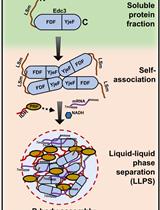
Assay to Study the Phase-transition Behavior of Edc3, a Conserved Processing Body (P-body) Marker Protein

研究保守的加工体 (P-body) 标记蛋白Edc3 相变行为的试验

RR Raju Roy
PR Purusharth I. Rajyaguru
2349 Views
Aug 20, 2022
RNA granules are conserved, non-membranous, biphasic structures predominantly composed of RNA and RNA-binding proteins. RNA granules often assemble as a result of cellular responses to a variety of stresses, including infection. Two types of RNA granules are best characterized: stress granules (SGs) and processing bodies (P-bodies). The mechanism of RNA granule assembly and disassembly is still understudied because of its complex composition and dynamic behavior. The assembly of RNA granules is driven by a process known as phase separation of granule components. Edc3 is a conserved decapping activator and an essential P-body component in Saccharomyces cerevisiae. Phase separation of P-body proteins has been poorly explored. This protocol will enable the visualization of the phase transition behavior of Edc3, since it is tagged to mCherry. It further describes using small molecules and other proteins to study P-body dynamics. In addition to the assembly of Edc3, this assay also lays the foundation to study disassembly of phase-separated assemblies in vitro, which was not explored earlier. We have devised the assay to describe the use of one such protein that acts as a disassembly factor. Overall, this protocol is simple to perform and can potentially be combined with analyzing these assemblies using other approaches.Graphical abstract:

生物工程

人诱导多能干细胞中人心肺祖细胞和微组织的共分化和共成熟

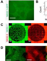
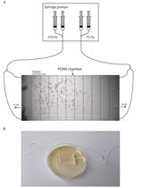
Co-differentiation and Co-maturation of Human Cardio-pulmonary Progenitors and Micro-Tissues from Human Induced Pluripotent Stem Cells

人诱导多能干细胞中人心肺祖细胞和微组织的共分化和共成熟

WN Wai Hoe Ng
BV Barbie Varghese
XR Xi Ren
2658 Views
Aug 20, 2022
Currently, there are several in vitro protocols that focus on directing human induced pluripotent stem cell (hiPSC) differentiation into either the cardiac or pulmonary lineage. However, these systemsprotocols are unable to recapitulate the critical exchange of signals and cells between the heart and lungs during early development. To address this gap, here we describe a protocol to co-differentiate cardiac and pulmonary progenitors within a single hiPSC culture by temporal specific modulation of Wnt and Nodal signaling. Subsequently, human cardio-pulmonary micro-tissues (μTs) can be generated by culturing the co-induced cardiac and pulmonary progenitors in 3D suspension culture. Anticipated results include expedited alveolarization in the presence of cardiac cells, and segregation of the cardiac and pulmonary μTs in the absence of exogenous Wnt signaling. This protocol can be used to model cardiac and pulmonary co-development, with potential applications in drug testing, and as a platform for expediting the maturation of pulmonary cells for lung tissue engineering.

癌症生物学

一种快速可靠的生成纯的、单细胞衍生的哺乳动物细胞克隆的方法

A Fast and Reliable Method to Generate Pure, Single Cell-derived Clones of Mammalian Cells

一种快速可靠的生成纯的、单细胞衍生的哺乳动物细胞克隆的方法

ZH Zhe Han
BM Bindhu K. Madhavan
SK Serap Kaymak
PN Peter Nawroth
VK Varun Kumar
3390 Views
Aug 20, 2022
Stable cell cloning is an essential aspect of biological research. All advanced genome editing tools rely heavily on stable, pure, single cell-derived clones of genetically engineered cells. For years, researchers have depended on single-cell dilutions seeded in 96- or 192-well plates, followed by microscopic exclusion of the wells seeded with more than or without a cell. This method is not just laborious, time-consuming, and uneconomical but also liable to unintentional error in identifying the wells seeded with a single cell. All these disadvantages may increase the time needed to generate a stable clone. Here, we report an easy-to-follow and straightforward method to conveniently create pure, stable clones in less than half the time traditionally required. Our approach utilizes cloning cylinders with non-toxic tissue-tek gel, commonly used for immobilizing tissues for sectioning, followed by trypsinization and screening of the genome-edited clones. Our approach uses minimal cell handling steps, thus decreasing the time invested in generating the pure clones effortlessly and economically.Graphical abstract: A schematic comparison showing the traditional dilution cloning and the method described here. Here, a well-separated colony (in the green box) must be preferred over the colonies not well separated (in the red box).
单克隆半高通量筛选的In-Cell Western实验方案

In-Cell Western Protocol for Semi-High-Throughput Screening of Single Clones

单克隆半高通量筛选的In-Cell Western实验方案

AP Arpita S. Pal
AA Alejandra M. Agredo
AK Andrea L. Kasinski
2935 Views
Aug 20, 2022
The in-cell western (ICW) is an immunocytochemical technique that has been used to screen for effects of siRNAs, drugs, and small molecule inhibitors. The reduced time and number of cells required to follow this protocol illustrates its semi-high-throughput nature. Performing a successful ICW protocol requires fixing and permeabilizing adherent cells directly in the plate that specifically exposes the epitope of interest. After blocking of non-specific proteins, the cells are incubated overnight with a primary antibody of interest, which is detected via a host-specific near-infrared fluorescently labeled LI-COR secondary antibody. In the final step, the plate is scanned using an Odyssey LI-COR Imaging System or similar, and each of the wells is quantified. For the first time, this technique has been demonstrated to be reproducibly utilized for semi-high-throughput selection of knockout or overexpression clones.Graphical abstract:

细胞生物学

用于急性肺损伤大型动物模型绿色组织病理学评价的半定量评分系统

A Semi-quantitative Scoring System for Green Histopathological Evaluation of Large Animal Models of Acute Lung Injury

用于急性肺损伤大型动物模型绿色组织病理学评价的半定量评分系统

IS Iran A. N. Silva
NG Nika Gvazava
DB Deniz A. Bölükbas
MS Martin Stenlo
JD Jiao Dong
SH Snejana Hyllen
LP Leif Pierre
SL Sandra Lindstedt
DW Darcy E. Wagner
3636 Views
Aug 20, 2022
Acute respiratory distress syndrome (ARDS) is a life-threatening, high mortality pulmonary condition characterized by acute lung injury (ALI) resulting in diffuse alveolar damage. Despite progress regarding the understanding of ARDS pathophysiology, there are presently no effective pharmacotherapies. Due to the complexity and multiorgan involvement typically associated with ARDS, animal models remain the most commonly used research tool for investigating potential new therapies. Experimental models of ALI/ARDS use different methods of injury to acutely induce lung damage in both small and large animals. These models have historically played an important role in the development of new clinical interventions, such as fluid therapy and the use of supportive mechanical ventilation (MV). However, failures in recent clinical trials have highlighted the potential inadequacy of small animal models due to major anatomical and physiological differences, as well as technical challenges associated with the use of clinical co-interventions [e.g., MV and extracorporeal membrane oxygenation (ECMO)]. Thus, there is a need for larger animal models of ALI/ARDS, to allow the incorporation of clinically relevant measurements and co-interventions, hopefully leading to improved rates of clinical translation. However, one of the main challenges in using large animal models of preclinical research is that fewer species-specific experimental tools and metrics are available for evaluating the extent of lung injury, as compared to rodent models. One of the most relevant indicators of ALI in all animal models is evidence of histological tissue damage, and while histological scoring systems exist for small animal models, these cannot frequently be readily applied to large animal models. Histological injury in these models differs due to the type and severity of the injury being modeled. Additionally, the incorporation of other clinical support devices such as MV and ECMO in large animal models can lead to further lung damage and appearance of features absent in the small animal models. Therefore, semi-quantitative histological scoring systems designed to evaluate tissue-level injury in large animal models of ALI/ARDS are needed. Here we describe a semi-quantitative scoring system to evaluate histological injury using a previously established porcine model of ALI via intratracheal and intravascular lipopolysaccharide (LPS) administration. Additionally, and owing to the higher number of samples generated from large animal models, we worked to implement a more sustainable and greener histopathological workflow throughout the entire process.
果蝇翅成虫芽单细胞悬浮液的制备

Preparation of a Single-cell Suspension from Drosophila Wing Imaginal Discs

果蝇翅成虫芽单细胞悬浮液的制备

SY Shu Yang
BS Brooke Sears
XZ Xiaoyan Zheng
2183 Views
Aug 20, 2022
The wing imaginal discs in Drosophila larvae are a pair of sac-like structures that later form the wings of the adult fly. During the past decades, wing discs have been used as a simple and accessible model system, for identifying genes and deciphering signaling cascades that play crucial roles in many aspects of development. In this protocol, we describe a simple method for preparing a cell suspension from wing discs (see Graphical abstract). This method can also be applied to the preparation of single-cell suspensions from other types of Drosophila tissues. When combined with genetic labeling, the dissociated cells are suitable for downstream analysis, such as flow cytometry. This method was recently used to isolate different populations of cells from Drosophila imaginal discs (Yang et al., 2022).Graphical abstract: Procedures to prepare a single-cell suspension from Drosophila imaginal discs. Illustration of the main steps to dissect, temporarily store, and dissociate imaginal discs from Drosophila larvae. Refer to the Procedure section for detailed description of each step.

免疫学

结合无菌损伤和微生物污染对小鼠术后腹膜粘连进行建模

Combination of Sterile Injury and Microbial Contamination to Model Post-surgical Peritoneal Adhesions in Mice

结合无菌损伤和微生物污染对小鼠术后腹膜粘连进行建模

JB Julia Bayer
DS Deborah Stroka
PK Paul Kubes
DC Daniel Candinas
JZ Joel Zindel
2283 Views
Aug 20, 2022
Abdominal surgeries are frequently associated with the development of post-surgical adhesions. These are irreversible fibrotic scar bands that appear between abdominal organs and the abdominal wall. Patients suffering from adhesions are at risk of severe complications, such as small bowel obstruction, chronic pelvic pain, or infertility. To date, no cure exists, and the understanding of underlying molecular mechanisms of adhesion formation is incomplete. The current paradigm largely relies on sterile injury mouse models. However, abdominal surgeries in human patients are rarely completely sterile procedures. Here, we describe a modular surgical procedure for simultaneous or separate induction of sterile injury and microbial contamination. Combined, these insults synergistically lead to adhesion formation in the mouse peritoneal cavity. Surgical trauma is confined to a localized sterile injury of the peritoneum. Microbial contamination of the peritoneal cavity is induced by a limited perforation of the microbe-rich large intestine or by injection of fecal content. The presented protocol extends previous injury-based adhesion models by an additional insult through microbial contamination, which may more adequately model the clinical context of abdominal surgery.Graphical abstract:

医学

通过低分辨率 SDS-琼脂糖凝胶电泳进行血友病因子多聚体分析

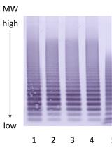
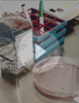
Von Willebrand Factor Multimer Analysis by Low Resolution SDS-Agarose Gel Electrophoresis

通过低分辨率 SDS-琼脂糖凝胶电泳进行血友病因子多聚体分析

HG Herbert Gritsch
MS Margit Stimpfl
PT Peter L. Turecek
2666 Views
Aug 20, 2022
Von Willebrand factor (VWF) is a complex glycoprotein found in plasma, composed of disulfide-bond-linked multimers with apparent molecular weights between 500 kDa and 20,000 kDa. After release of VWF from storage granules, it is cleaved in flowing blood by the specific metalloproteinase ADAMTS13, resulting in a highly characteristic cleavage pattern and structure. As the structure of VWF multimers determines diagnosis of von Willebrand disease, which has different sub-types with different multimer- and cleavage patterns, VWF analysis is performed using low-resolution horizontal SDS-agarose gel electrophoresis. However, almost every laboratory uses a different protocol, and all experimental details are rarely, if at all, described. Therefore, the results from similar methods may be substantially different. Here, we present a detailed description of a validated VWF multimer method that we have developed. It has been successfully used for over more than 20 years in quality control of recombinant and plasma-derived VWF drug products, and in preclinical and clinical studies with VWF drug candidates. As most of the published methods, it enables visualization of VWF multimers separated by electrophoresis by immunostaining with a polyclonal anti-human VWF antibody followed by a secondary antibody coupled to alkaline phosphatase. VWF appears as a series of regularly spaced bands on the low and middle molecular weight range of the gel, with an unresolved zone in the high molecular weight (HMW) range, where ultra-large multimers are found. An example is shown below. This low-resolution agarose gel electrophoresis allows the determination of the number of VWF multimers with high reproducibility.Graphical abstract: Example of electrophoretic analysis of multimer structure of four batches of a recombinant VWF drug substance.

神经科学

秀丽隐杆线虫的趋气性测定以研究行为可塑性

Aerotaxis Assay in Caenorhabditis elegans to Study Behavioral Plasticity

秀丽隐杆线虫的趋气性测定以研究行为可塑性

QL Qiaochu Li
DM Daniel-Cosmin Marcu
PD Paul H. Dear
KB Karl Emanuel Busch
1761 Views
Aug 20, 2022
C. elegans shows robust and reproducible behavioral responses to oxygen. Specifically, worms prefer O2 levels of 5–10% and avoid too high or too low O2. Their O2 preference is not fixed but shows plasticity depending on experience, context, or genetic background. We recently showed that this experience-dependent plasticity declines with age, providing a useful behavioral readout for studying the mechanisms of age-related decline of neural plasticity. Here, we describe a technique to visualize behavioral O2 preference and its plasticity in C. elegans, by creating spatial gradients of [O2] in a microfluidic polydimethylsiloxane (PDMS) chamber and recording the resulting spatial distribution of the animals.

植物科学

本氏烟草的瞬态系统进行荧光标记 ATG8 的共定位分析

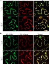
Colocalization Assay with Fluorescent-tagged ATG8 Using a Nicotiana benthamiana-based Transient System

本氏烟草的瞬态系统进行荧光标记 ATG8 的共定位分析

JM Jinyan Mai
DS Dandan Shang
FL Faqiang Li
NL Na Luo
2699 Views
Aug 20, 2022
Autophagy is an evolutionarily conserved intracellular degradation process. During autophagy, a set of autophagy-related (ATG) proteins orchestrate the formation of double-bound membrane vesicles called autophagosomes to engulf cytoplasmic material and deliver it to the vacuole for breakdown. Among ATG proteins, the ATG8 is the only one decorating mature autophagosomes and therefore is regarded as a bona fide autophagic marker; colocalization assays with ATG8 are wildly used as a reliable method to identify the components of autophagy machinery or autophagic substrates. Here, we describe a colocalization assay with fluorescent-tagged ATG8 using a tobacco (Nicotiana benthamiana)-based transient expression system.