往期刊物2020

卷册: 10, 期号: 10

left

Dec

20

Dec

5

Nov

20

Nov

5

Oct

20

Oct

5

Sep

20

Sep

5

Aug

20

Aug

5

Jul

20

Jul

5

Jun

20

Jun

5

May

20

May

5

Apr

20

Apr

5

Mar

20

Mar

5

Feb

20

Feb

5

Jan

20

Jan

5

right

生物化学

细菌膜蛋白在蛋白质脂质体中的体外整合

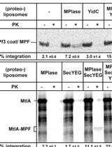
In vitro Assay for Bacterial Membrane Protein Integration into Proteoliposomes

细菌膜蛋白在蛋白质脂质体中的体外整合

HN Hanako Nishikawa
MS Masaru Sasaki
KN Ken-ichi Nishiyama
4920 Views
May 20, 2020
It is important to experimentally determine how membrane proteins are integrated into biomembranes to unveil the roles of the integration factors, and to understand the functions and structures of membrane proteins. We have developed a reconstitution system for membrane protein integration in E. coli using purified factors, in which the integration reaction in vivo is highly reproducible. This system enabled not only analysis of membrane-embedded factors including glycolipid MPIase, but also elucidation of the detailed mechanisms underlying membrane protein integration. Using the system, the integration of membrane proteins can be evaluated in vitro through a protease-protection assay. We report here how to prepare (proteo)liposomes and to determine the activities of membrane protein integration.
通过移码荧光蛋白报告基因评估基因组编辑工具的效率

Evaluation of the Efficiency of Genome Editing Tools by a Frameshift Fluorescence Protein Reporter

通过移码荧光蛋白报告基因评估基因组编辑工具的效率

BM Balaji T. Moorthy
Akhilesh  Kumar Akhilesh Kumar
LL Lauren X. Lotenfoe
FZ Fangliang Zhang
4457 Views
May 20, 2020
In the last decade, genome editing has been the center of attention as a novel tool for mechanistic investigations and for potential clinical applications. Various genome editing tools like meganucleases, zinc finger nucleases (ZFNs), transcription activator-like effector-based nucleases (TALEN), and the clustered regularly interspaced short palindromic repeats (CRISPR)-associated genes (Cas), have been developed in recent years. For the optimal use as well as continued developments of these genome editing tools, the evaluation of their efficiencies and accuracies is vital. Here, we present a protocol for a reporter based on frameshift fluorescence protein which we recently developed to evaluate the efficiency and accuracy of genome editing tools. In this method, a ~20 bp target sequence containing frame-shifting is inserted after the start codon of a cerulean fluorescence protein (CFP) to inactivate its fluorescence, and only a new insertion/deletion event in the target sequence will reactivate the CFP fluorescence. To increase the traceability, an internal ribosome entry site and a red fluorescence protein, mCherryFP, are placed downstream of the reporter. The percentage of CFP-positive cells resulted from in/del mediated fluorescence restoration can be quantified by fluorescence measuring devices as the readout for genome editing frequency. As a demonstration, we present the usage for CRISPR-Cas9 technique here with flow cytometer as the readout for fluorescence changes.
硫酸多糖的全甲基化和微萃取质谱分析

Permethylation and Microfractionation of Sulfated Glycans for MS Analysis

硫酸多糖的全甲基化和微萃取质谱分析

SY Shin-Yi Yu
SS Sergei Snovida
KK Kay-Hooi Khoo
3977 Views
May 20, 2020
Sulfated glycans are barely detectable in routine mass spectrometry (MS)-based glycomic analysis due to ion suppression by the significantly more abundant neutral glycans in the positive ion mode, and sialylated non-sulfated glycans in the negative ion mode, respectively. Nevertheless, the negative charge imparted by sulfate can be advantageous for selective detection in the negative ion mode if the sialic acids can first be neutralized. This is most conveniently achieved by a concerted sample preparation workflow in which permethylation is followed by solid phase fractionation to isolate the sulfated glycans prior to MS analysis. Importantly, we demonstrated that conventional NaOH/DMSO slurry permethylation method can retain the sulfates. Instead of extracting permethylated glycans into chloroform for sample clean-up, reverse phase C18 cartridge coupled with self-packed amine-tip or mixed mode weak anion exchange cartridge can be utilized to obtain in good yield the non-sulfated, mono-sulfated, and multiply sulfated permethylated glycans in separate fractions for sulfoglycomic analysis.
甲基化硫酸多糖的负离子模式nanoLC-ESI-MS / MS分析

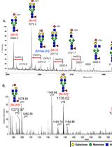
Negative Ion Mode nanoLC-ESI-MS/MS Analyses of Permethylated Sulfated Glycans

甲基化硫酸多糖的负离子模式nanoLC-ESI-MS / MS分析

SY Shin-Yi Yu
CC Chu-Wen Cheng
KK Kay-Hooi Khoo
3868 Views
May 20, 2020
We have developed enabling techniques for sulfoglycomics based on MALDI-MS mapping and MS/MS sequencing of permethylated sulfated glycans. We then extended further the analytical workflow to C18 reverse phase (RP)-nanoLC-nanoESI-MS/MS analyses of permethylated sulfated glycans in the negative ion mode. The advantages are that extra sulfates on permethylated di- and multiply sulfated glycans will survive in nanoESI conditions to allow detection of multiply charged intact molecular ions, and more comprehensive MS/MS can be performed in an automated fashion at higher sensitivity, compared with MALDI-MS/MS. Parallel higher energy collision dissociation (HCD) and ion trap collision induced dissociation (CID)-based MS2, coupled with product-dependent MS3 in data dependent acquisition mode proved to be highly productive when applied to resolve and identify the isomeric sulfated glycan structures. In-house glycomic data mining software, GlyPick, was developed and used to automate the downstream process of identification and relative quantification of target sulfated glycotopes based on summed intensity of their diagnostic MS2 ions extracted from thousands of HCD-MS2 and/or CID-MS2 data.

癌症生物学

NOTCH1诱导小鼠T细胞急性淋巴细胞白血病模型的建立

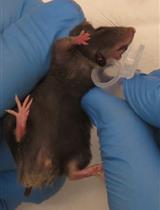
Modeling NOTCH1 driven T-cell Acute Lymphoblastic Leukemia in Mice

NOTCH1诱导小鼠T细胞急性淋巴细胞白血病模型的建立

AW Agnieszka A. Wendorff
AF Adolfo A. Ferrando
5746 Views
May 20, 2020
T-cell acute lymphoblastic leukemia (T-ALL) is an aggressive hematological malignancy that arises from transformation of T-cell primed hematopoietic progenitors. Although T-ALL is a heterogenous and molecularly complex disease, more than 65% of T-ALL patients carry activating mutations in the NOTCH1 gene. The majority of T-ALL–associated NOTCH1 mutations either disrupt the negative regulatory region, allowing signal activation in the absence of ligand binding, or result in truncation of the C-terminal PEST domain involved in the termination of NOTCH1 signaling by proteasomal degradation. To date, retroviral transduction models have relied heavily on the overexpression of aggressively truncated variants of NOTCH1 (such as ICN1 or ΔE-NOTCH1), which result in supraphysiological levels of signaling activity and are rarely found in human T-ALL. The current protocol describes the method for mouse bone marrow isolation, hematopoietic stem and progenitor cell (HSC) enrichment, followed by retroviral transduction with an oncogenic mutant form of the NOTCH1 receptor (NOTCH1-L1601P-ΔP) that closely resembles the gain-of-function mutations most commonly found in patient samples. A hallmark of this forced expression of constitutively active NOTCH1 is a transient wave of extrathymic immature T-cell development, which precedes oncogenic transformation to T-ALL. Furthermore, this approach models leukemic transformation and progression in vivo by allowing for crosstalk between leukemia cells and the microenvironment, an aspect unaccounted for in cell-line based in vitro studies. Thus, the HSC transduction and transplantation model more faithfully recapitulates development of the human disease, providing a highly comprehensive and versatile tool for further in vivo and ex vivo functional studies.
基于与血管距离的肿瘤细胞分离

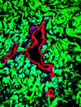
Isolation of Tumor Cells Based on Their Distance from Blood Vessels

基于与血管距离的肿瘤细胞分离

SK Saran Kumar
HS Husni Sharife
TK Tirzah Kreisel
LB Libat Bar-Lev
MG Myriam Grunewald
EK Eli Keshet
5157 Views
May 20, 2020
Differential exposure of tumor cells to microenvironmental cues greatly impacts cell phenotypes, raising a need for position based sorting of tumor cells amenable to multiple OMICs and functional analyses. One such key determinant of tumor heterogeneity in solid tumors is its vasculature. Proximity to blood vessels (BVs) profoundly affects tumor cell phenotypes due to differential availability of oxygen, gradient exposure to blood-borne substances and inputs by angiocrine factors. To unravel the whole spectrum of genes, pathways and phenotypes impacted by BVs and to determine spatial domains of vascular influences, we developed a methodology for sorting tumor cells according to their relative distance from BVs. The procedure exemplified here using glioblastoma (GBM) model is based on differential uptake of intra-venously injected, freely-diffusing fluorescent dye that allows separation of stroma-free tumor cells residing in different, successive microenvironments amenable for subsequent OMICs and functional analyses. This reliable, easy to use, cost effective strategy can be extended to all solid tumors to study the impact of vasculature or the lack of it.

细胞生物学

Masson Trichrome染色法分析小鼠组织切片中的普遍纤维化

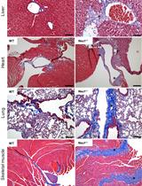
Analysis of Generalized Fibrosis in Mouse Tissue Sections with Masson’s Trichrome Staining

Masson Trichrome染色法分析小鼠组织切片中的普遍纤维化

Dv Diantha van de Vlekkert
EM Eda Machado
Ad Alessandra d’Azzo
8995 Views
May 20, 2020
Expansion of fibrous connective tissue and abnormal deposition of extracellular matrix (ECM) are at the basis of many fibrotic diseases. Fibrosis can occur in response to both physiological and pathological cues, including wound healing, tissue remodeling/repair and inflammation. Chronic fibrosis can lead to severe tissue damage, organ failure and death. Assessing the extent of organ fibrosis is crucial for accurate diagnosis of this condition. The use of Masson’s trichrome staining of tissue sections from skeletal muscle is a fast method for detection of morphological alterations indicative of a fibrotic phenotype in this organ. This staining method detects the extent of collagen fibers deposition and, because it employs the combination of three dyes, can also distinguish muscle fibers (red), from collagen (blue) and nuclei (black), simultaneously.
用于耗氧量和乳酸生产分析的胰腺腺泡细胞制备

Pancreatic Acinar Cell Preparation for Oxygen Consumption and Lactate Production Analysis

用于耗氧量和乳酸生产分析的胰腺腺泡细胞制备

JA Jane A. Armstrong
RS Robert Sutton
DC David N. Criddle
4190 Views
May 20, 2020
Mitochondrial dysfunction is a principal feature of acute pancreatitis (AP) although the underlying mechanisms are still unclear. AP precipitants induce Ca2+-dependent formation of the mitochondrial permeability transition pore (MPTP) in pancreatic acinar cells (PACs), leading to ATP depletion and necrosis. Evaluations of mitochondrial bioenergetics have mainly been performed in isolated PACs using confocal microscopy, with assessment of mitochondrial membrane potential, NADH/FAD+ and ATP levels, coupled with patch-clamp electrophysiology. These studies are technically demanding and time-consuming. Application of Seahorse flux analysis now allows detailed investigations of bioenergetics changes to be performed in cell populations using a multi-well plate-reader format; rates of oxygen consumption (OCR) and extracellular acidification (ECAR) provide important information about cellular respiration and glycolysis, respectively. Parameters such as maximal respiration, ATP-linked capacity and proton leak can be derived from application of a respiratory function “stress” test that involves pharmacological manipulation of the electron transport chain. The use of Seahorse Flux analysis therefore provides a quick, and convenient means to measure detailed cellular bioenergetics and allows results to be coupled with other plate-reader based assays, providing a fuller understanding of the pathophysiological consequences of mitochondrial bioenergetics alterations.
空间图像相关光谱法(ICS):一种从荧光显微图像中确定亚细胞聚集结构平均尺寸的技术

Spatial Image Correlation Spectroscopy (ICS): A Technique for Average Size Determination of Subcellular Accumulated Structures from Fluorescence Microscopic Images

空间图像相关光谱法(ICS):一种从荧光显微图像中确定亚细胞聚集结构平均尺寸的技术

AK Akira Kitamura
MK Masakata Kinjo
3930 Views
May 20, 2020
Size determination of subcellular structures such as inclusion bodies (IBs) and granules from fluorescent images is important for identification and structural characterization. However, it is often time-consuming just for the comparison of the average size of the structures. Here, we introduce a high-throughput procedure to represent the average size of structures in fluorescent images using Spatial Image Correlation Spectroscopy (SICS). This procedure provides an easier comparison of bodies and granular structures such as inclusion bodies (IBs) including misfolded protein aggregation, granules containing RNA (e.g., stress granules and processing bodies).

发育生物学

转录因子强化表达直接将小鼠胚胎成纤维细胞重编程为常规1型树突状细胞

Direct Reprogramming of Mouse Embryonic Fibroblasts to Conventional Type 1 Dendritic Cells by Enforced Expression of Transcription Factors

转录因子强化表达直接将小鼠胚胎成纤维细胞重编程为常规1型树突状细胞

FR Fábio F. Rosa
CP Cristiana F. Pires
OZ Olga Zimmermannova
CP Carlos-Filipe Pereira
6578 Views
May 20, 2020
Ectopic expression of transcription factor combinations has been recently demonstrated to reprogram differentiated somatic cells towards the dendritic cell (DC) lineage without reversion to a multipotent state. DCs have the ability to induce potent and long-lasting adaptive immune responses. In particular, conventional type 1 DCs (cDC1s) excel on antigen cross-presentation, a critical step for inducing CD8+ T cell cytotoxic responses. The rarity of naturally occurring cDC1s and lack of in vitro methodologies for the generation of pure cDC1 populations strongly hinders the study of cDC1 lineage specification and function. Here, we describe a protocol for the generation of induced DCs (iDCs) by lentiviral-mediated expression of the transcription factors PU.1, IRF8 and BATF3 in mouse embryonic fibroblasts. iDCs acquire DC morphology, cDC1 phenotype and transcriptional signatures within 9 days. iDCs generated with this protocol acquire functional ability to respond to inflammatory stimuli, engulf dead cells, process and cross-present antigens to CD8+ T cells. DC reprogramming provides a simple and tractable system to generate high numbers of cDC1-like cells for high content screening, opening new avenues to better understand cDC1 specification and function. In the future, faithful induction of cDC1 fate in fibroblasts may lead to the generation of patient-specific DCs for vaccination.

免疫学

结核分枝杆菌感染小鼠肺和支气管肺泡灌洗样品中髓样细胞的流式细胞仪分析和荧光激活细胞分选

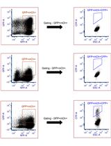
Flow Cytometry Analysis and Fluorescence-activated Cell Sorting of Myeloid Cells from Lung and Bronchoalveolar Lavage Samples from Mycobacterium tuberculosis-infected Mice

结核分枝杆菌感染小鼠肺和支气管肺泡灌洗样品中髓样细胞的流式细胞仪分析和荧光激活细胞分选

Alissa C. Rothchild Alissa C. Rothchild
DM Dat Mai
AA Alan Aderem
AD Alan H. Diercks
6178 Views
May 20, 2020
Mycobacterium tuberculosis (Mtb) is transmitted by aerosol and can cause serious bacterial infection in the lung that can be fatal if left untreated. Mtb is now the leading cause of death worldwide by an infectious agent. Characterizing the early events of in vivo infection following aerosol challenge is critical for understanding how innate immune cells respond to infection but is technically challenging due to the small number of bacteria that initially infect the lung. Previous studies either evaluated Mtb-infected cells at later stages of infection when the number of bacteria in the lung is much higher or used in vitro model systems to assess the response of myeloid cells to Mtb. Here, we describe a method that uses fluorescent bacteria, a high-dose aerosol infection model, and flow cytometry to track Mtb-infected cells in the lung immediately following aerosol infection and fluorescence-activated cell sorting (FACS) to isolate naïve, bystander, and Mtb-infected cells for downstream applications, including RNA-sequencing. This protocol provides the ability to monitor Mtb-infection and cell-specific responses within the context of the lung environment, which is known to modulate the function of both resident and recruited populations. Using this protocol, we discovered that alveolar macrophages respond to Mtb infection in vivo by up-regulating a cell protective transcriptional response that is regulated by the transcription factor Nrf2 and is detrimental to early control of the bacteria.

微生物学

秀丽隐杆线虫的细菌草坪趋避和细菌双选择参照实验

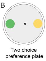
Bacterial Lawn Avoidance and Bacterial Two Choice Preference Assays in Caenorhabditis elegans

秀丽隐杆线虫的细菌草坪趋避和细菌双选择参照实验

JS Jogender Singh
AA Alejandro Aballay
4706 Views
May 20, 2020
Physical avoidance of pathogens is a crucial defense strategy used by the host to reduce pathogen infection. Hosts display the use of multiple strategies to sense and avoid pathogens, ranging from olfaction to sensing of damage caused by pathogen infection. Understanding various mechanisms of pathogen avoidance has the potential to uncover conserved host defense responses that are important against pathogen infections. Here, we describe protocols for studying pathogen lawn avoidance behavior as well as a change of bacterial preferences in the model nematode Caenorhabditis elegans. Besides, we describe the protocol for measuring preferences for pathogenic and nonpathogenic bacteria after training of the animals on pathogenic bacteria. These assays can be implemented in discovering various mechanisms of host learning that result in the avoidance of pathogens.

分子生物学

重组输入蛋白-组蛋白配合物分子大小的超离心分析

Molecular Size Analysis of Recombinant Importin-histone Complexes Using Analytical Ultracentrifugation

重组输入蛋白-组蛋白配合物分子大小的超离心分析

AP Abhilash Padavannil
CB Chad A. Brautigam
YC Yuh Min Chook
3882 Views
May 20, 2020
Histones constitute the protein components of nucleosomes. Despite their small sizes, histones do not diffuse through the nuclear pore complex. Instead, they are transported to the nucleus by importins, either alone or in complex with histone chaperones. Determining the molecular size of the importin-histone complexes is key to understanding the mechanism of histone transport and also the potential roles of importins as histone chaperones and in the assembly of nucleosomes. Here we report a simple and reproducible sedimentation-velocity based method to determine the molecular sizes of importin-histone complexes using analytical ultracentrifugation. The method does not use any reporter tags or interaction with column resin thereby analyzing the interactions of the native proteins.

神经科学

果蝇IBMPFD模型的建立、分析及体内药物治疗

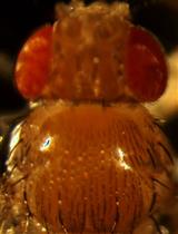
Generation, Analyzing and in-vivo Drug Treatment of Drosophila Models with IBMPFD

果蝇IBMPFD模型的建立、分析及体内药物治疗

TZ Ting Zhang
BH Bruce A. Hay
MG Ming Guo
4494 Views
May 20, 2020
Missense mutations of p97/cdc48/Valosin-containing protein (VCP) cause inclusion body myopathy, Paget disease with frontotemporal dementia (IBMPFD) and other neurodegenerative diseases. The pathological mechanism of IBMPFD is not clear and there is no treatment. We generated Drosophila models of IBMPFD in adult flight muscle in vivo. Here we describe a variety of assays to characterize disease pathology and dissect disease mechanism, and the consequences of in vivo feeding of VCP inhibitors.

植物科学

 Cyanobacterium Synechocystis sp. PCC 6803 中蛋白混合物的标记亲和层析纯化方法

Purification of Protein-complexes from the Cyanobacterium Synechocystis sp. PCC 6803 Using FLAG-affinity Chromatography

Cyanobacterium Synechocystis sp. PCC 6803 中蛋白混合物的标记亲和层析纯化方法

MK Minna M. Koskela
PS Petra Skotnicová
ÉK Éva Kiss
RS Roman Sobotka
5194 Views
May 20, 2020
Exploring the structure and function of protein complexes requires their isolation in the native state–a task that is made challenging when studying labile and/or low abundant complexes. The difficulties in preparing membrane-protein complexes are especially notorious. The cyanobacterium Synechocystis sp. PCC 6803 is a widely used model organism for the physiology of oxygenic phototrophs, and the biogenesis of membrane-bound photosynthetic complexes has traditionally been studied using this cyanobacterium. In a typical approach, the protein complexes are purified with a combination of His-affinity chromatography and a size-based fractionation method such as gradient ultracentrifugation and/or native electrophoresis. However, His-affinity purification harbors prominent contaminants and the levels of many proteins are too low for a feasible multi-step purification. Here, we have developed a purification method for the isolation of 3x FLAG-tagged proteins from the membrane and soluble fractions of Synechocystis. Soluble proteins or solubilized thylakoids are subjected to a single affinity purification step that utilizes the highly specific binding of FLAG-affinity resin. After an intensive wash, the captured proteins are released from the resin under native conditions using an excess of synthetic 3x FLAG peptide. The protocol allows fast isolation of low abundant protein complexes with a superb purity.