往期刊物2019

卷册: 9, 期号: 15

left

Dec

20

Dec

5

Nov

20

Nov

5

Oct

20

Oct

5

Sep

20

Sep

5

Aug

20

Aug

5

Jul

20

Jul

5

Jun

20

Jun

5

May

20

May

5

Apr

20

Apr

5

Mar

20

Mar

5

Feb

20

Feb

5

Jan

20

Jan

5

right

癌症生物学

卵巢癌3D模型中的光动力学治疗

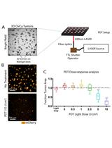
Photodynamic Therapy in a 3D Model of Ovarian Cancer

卵巢癌3D模型中的光动力学治疗

SN Shubhankar Nath
KM Kaitlin Moore
4926 Views
Aug 5, 2019
Photodynamic therapy (PDT), is a clinically-approved light-based anti-cancer treatment modality in which a photoactivatable photosensitizer is irradiated with an appropriate wavelength of light to generate cytotoxic molecules to kill cancer cells. In this article, we describe an in vitro PDT protocol using a 3-dimensional (3D) model of ovarian cancer that was established on beds of Matrigel. PDT was performed using a liposomal formulation of verteporfin photosensitizer (Visudyne®). The cancer cells were genetically-labeled with the fluorescent protein mCherry to facilitate the evaluation of the treatment response. This protocol is advantageous because the mCherry fluorescence is an indicator of cell viability, eliminating the need for external dyes, which often exhibit limited penetration and diffusion into 3D organoids. Additionally, Visudyne PDT achieves significant tumor-killing efficacy in a 3D model for ovarian cancer.

细胞生物学

发芽酵母和裂殖酵母中基于QUEEN的时空ATP成像

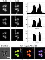
QUEEN-based Spatiotemporal ATP Imaging in Budding and Fission Yeast

发芽酵母和裂殖酵母中基于QUEEN的时空ATP成像

MT Masak Takaine
6133 Views
Aug 5, 2019
Yeasts have provided an exceptional model for studying metabolism and bioenergetics in eukaryotic cells. Among numerous metabolites, adenosine triphosphate (ATP) is a major metabolite that is essential for all living organisms. Therefore, a clearer understanding of ATP dynamics in living yeast cells is important for deciphering cellular energy metabolism. However, none of the methods currently available to measure ATP, including biochemical analyses and ATP indicators, have been suitable for close examinations of ATP concentrations in yeast cells at the single cell level. Using the recently developed ATP biosensor QUEEN, which is suitable for yeasts and bacteria, a protocol was described herein to visualize ATP concentrations in living budding and fission yeast cells. This simple method enables the easy and reliable examination of ATP dynamics in various yeast mutants, thereby providing novel molecular insights into cellular energy metabolism.

免疫学

脾切除术限制小鼠应激反应的精准技术用于精确现实检测炎症和免疫

Precision Technique for Splenectomy Limits Mouse Stress Responses for Accurate and Realistic Measurements for Investigating Inflammation and Immunity

脾切除术限制小鼠应激反应的精准技术用于精确现实检测炎症和免疫

SL Shengwen Calvin Li
AR Anthony D. Rangel
MK Mustafa H. Kabeer
5575 Views
Aug 5, 2019
Splenectomy in an animal model requires a standardized technique utilizing best practice to avoid variability which can result in adverse impact to the animal resulting in flawed physiologic responses simply due to technique rather than to the studied variables. In the case of the spleen, often investigators are analyzing the animal immune or inflammatory responses. Surgical splenectomy involves many variables from the training and expertise of the surgeon, which directly correlates to surgical technique to the length of operation and ease of the procedure. This operation, in turn, impacts blood loss and insensible fluid losses, sterile technique, unintended trauma to the spleen and surrounding organs, the length of the incision and the duration of the operation with more prolonged exposure to anesthetic agents. All these variables ultimately play a significant role in the experiment since they directly affect the response of the model in terms of inflammation, immune activation, or even suppression. Undesired variables such as these go unnoticed and lead to inaccurate and misleading data.
用活体生物荧光法观察小鼠白念珠菌感染模型中的缺氧情况

Visualizing Hypoxia in a Murine Model of Candida albicans Infection Using in vivo Biofluorencence

用活体生物荧光法观察小鼠白念珠菌感染模型中的缺氧情况

José Pedro Lopes José Pedro Lopes
CU Constantin F. Urban
4482 Views
Aug 5, 2019
Candida albicans is a leading human fungal pathogen that uses several metabolic adaptations to escape immune cells and causes systemic disease. Here, we describe a protocol for measuring one of these adaptations, the ability to thrive in hypoxic niches. Hypoxia was generated after successful subdermal infection with C. albicans in a murine infection model. Hypoxia was measured using a fluorescent dye for carbonic anhydrase 9, a host enzyme active under hypoxic conditions. Emitted fluorescence was subsequently quantified using an IVIS system. This protocol was optimized for the use in subdermal infection in mice but has the potential to be adapted to other models of fungal infection.

微生物学

FDAA用于肽聚糖原位标记的优选方案

Optimized Protocol for the Incorporation of FDAA (HADA Labeling) for in situ Labeling of Peptidoglycan

FDAA用于肽聚糖原位标记的优选方案

KP Katharina Peters
MP Manuel Pazos
MV Michael S. VanNieuwenhze
WV Waldemar Vollmer
8011 Views
Aug 5, 2019
The essential peptidoglycan (PG) layer surrounds the cytoplasmic membrane in nearly all bacteria. It is needed to maintain the shape of the cell and protect it from lysis due to high turgor. Growth of the PG layer is a complex process that involves the activities of PG synthases and hydrolases during elongation and cell division. PG growth sites can be labeled by the recently developed fluorescent D-amino acid (FDAA) probes in a range of different bacteria. FDAAs are incorporated into PG by DD-transpeptidases (Penicillin-binding proteins, PBPs) or, if present, LD-transpeptidase (LDTs). Long-pulse in situ labeling of E. coli cells with the FDAA 7-hydroxycoumarincarbonylamino-D-alanine (HADA) is expected to result in a uniform label at the side wall of cells and enhanced label at cell division sites due to the intense PG synthesis. However, we observed reduced label at mid-cell when labeling E. coli cells with HADA. We reasoned that probe incorporated at cell division sites may be removed by PG hydrolases and modified the labeling protocol to better preserve PG-incorporated HADA for fluorescence microscopy. Here, we report the optimized HADA-labeling protocol by which cells retain an enhanced HADA signal at the division septum.
大肠杆菌特异性单链DNA测序(4S-seq)

Strand-specific Single-stranded DNA Sequencing (4S-seq) of E. coli genomes

大肠杆菌特异性单链DNA测序(4S-seq)

TM Takahiro Masuda
NK Nobuaki Kono
MT Masaru Tomita
KA Kazuharu Arakawa
6887 Views
Aug 5, 2019
Most bacterial genomes have biased nucleotide composition, and the asymmetry is considered to be caused by a single-stranded DNA (ssDNA) deamination arising from the bacterial replication machinery. In order to evaluate the relationship experimentally, the position and frequency of ssDNA formed during replication must be verified clearly. Although many ssDNA detection technologies exist, almost all methods have been developed for eukaryotic genomes. To apply these to bacterial genomes, which harbor a smaller amount of DNA than those of eukaryotes, more efficient, new methods are required. Therefore, we developed a novel strand-specific ssDNA sequencing method, called 4S-seq, for the bacterial genome. The 4S-seq method enriches ssDNA in the extracted genomic DNA by a dsDNA-specific nuclease and implements a strand-specific library using a biotin label with a customized tag. As a result, the 4S-seq is able to calculate the ssDNA content in each strand (Watson/Crick) at each position of the genome efficiently.
用于病原体检测的三维显微组织列阵中固相PCR

Solid Phase PCR on 3D Microstructure ArrayChip for Pathogen Detection Application

用于病原体检测的三维显微组织列阵中固相PCR

KK Krishna Kant
TN Tien Anh Ngo
5647 Views
Aug 5, 2019
Advanced free angle photolithography (FAPL) is presented for making 3D supercritical angle fluorescence (SAF) microstructures and transfer them on to polymeric chips using injection molding technique for low-cost microfluidic devices embedded with optical sensing structures. A solid phase polymerase chain reaction (SP-PCR) is used as model technique, which allows rapid and sensitive detection of pathogen DNA on-chip. This article presents the detailed fabrication of SAF structure and SP-PCR application on SAF structure for pathogen detection. This protocol of developing SAF structures using the FAPL process, increases the number of SAF per mm2. FAPL was performed via a motorized stage to control the angle of incidence and to achieve the desired bucket-shapes (dimensions of 50 μm to 150 μm with a slope) required for the 3D optical sensing. Due to the unique properties of SAF structures, it enhances the fluorescent signal by 46 times. Increasing the number of SAF structures and reducing the size resulted in reduction of sample volume required per test along with improvement in the limit of detection (LOD) due to a smaller size. This article also presents the experimental details of SP-PCR using DNA oligos bound to the SAF structures for on-chip pathogen detection and a comparison between different sizes of SAF structures. The direct on-chip SP-PCR paves the path for the application of this technique in point-of-care devices.
基于成像量化分析牛病毒性腹泻病毒的细胞间传播

Image-based Quantification of Direct Cell-to-cell Transmission of Bovine Viral Diarrhea Virus

基于成像量化分析牛病毒性腹泻病毒的细胞间传播

FM Fernando Merwaiss
DA Diego E. Alvarez
4639 Views
Aug 5, 2019
Different viruses rely on direct cell-to-cell transmission to propagate infection within the infected host. Measuring this mode of transmission in cultured cells is often complicated by the contribution of cell free viruses to spread, and the difficulty to distinguish between primary infected cells that produce the virus and neighboring cells that are the target of spreading. Here, we present a protocol to quantify cell-to-cell transmission of the model pestivirus bovine viral diarrhea virus that is based on the co-culture of producer cells that are infected with a reporter virus expressing mCherry and target cells that stably express GFP. Spread of cell-free viruses is blocked by the presence of a neutralizing antibody in the cell culture medium, and cell-associated transmission is unequivocally quantified by numbering cells that are positive for both GFP and mCherry using automated analysis of fluorescence microscopy images.
稻瘟病菌内吞作用的检测

Endocytosis Detection in Magnaporthe oryzae

稻瘟病菌内吞作用的检测

ML Muxing Liu
Zhengguang  Zhang Zhengguang Zhang
4149 Views
Aug 5, 2019
Endocytosis is an intracellular trafficking pathway that occurs in nutrient uptake, signal transduction and reconstruction of cell polarity and is conserved in eukaryotic cells. In fungi, endocytosis plays crucial roles in the physiology of hyphal growth and pathogenicity. vidence for endocytosis in filamentous fungi is detected by the membrane-selective dyes FM4-64. Cells of a range of filamentous fungal species readily take up these dyes. However, the method for endocytosis detection has not been well established in Magnaporthe oryzae. Here, we provide a protocol for tracking endocytosis in Magnaporthe oryzae.

分子生物学

秀丽隐杆线虫细胞特异性mRNA的核糖体亲和纯化

Cell Type-specific mRNA Purification in Caenorhabditis elegans via Translating Ribosome Affinity Purification

秀丽隐杆线虫细胞特异性mRNA的核糖体亲和纯化

IM Ian G. McLachlan
SF Steven W. Flavell
5102 Views
Aug 5, 2019
Cell type-specific molecular profiling is widely used to gain new insights into the diverse cell types that make up complex biological tissues. Translating ribosome affinity purification (TRAP) is a method in which the cell type-specific expression of epitope-tagged ribosomal subunits allows one to purify actively translating mRNAs without the need for cell sorting or fixation. We adapted this method for use in C. elegans to identify novel transcripts in single cell types or to identify the effects of environmental changes on the transcriptomes of larger cohorts of cells. In this protocol, we describe methods to generate transgenic animals bearing tagged ribosomes in cells of interest, prepare these animals for immunoprecipitation, purify ribosome-mRNA complexes, and obtain purified mRNA for next-generation sequencing.
利用改进的宿主细胞再活化反应进行人外周血单核细胞的DNA修复能力的离体检测

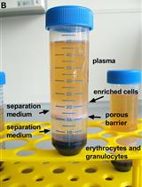
Ex vivo Analysis of DNA Repair Capacity of Human Peripheral Blood Mononuclear Cells by a Modified Host Cell Reactivation Assay

利用改进的宿主细胞再活化反应进行人外周血单核细胞的DNA修复能力的离体检测

KM Katja Matt
JB Jörg Bergemann
4224 Views
Aug 5, 2019
The ability of humans to repair DNA damages decreases with increasing age. In order to be able to repair daily occurring DNA damages, it becomes more and more important to preserve repair capability of cells with aging. The preservation of DNA repair processes contributes to preventing DNA mutations and subsequently the onset of age-related diseases such as cancer. For the determination of DNA repair of human cells, mostly in vitro cell cultures are used. However, an ex vivo approach can provide a more accurate result compared with in vitro cell cultures, since the DNA repair ability is measured directly without the influence of prolonged culture time. Published protocols use in vitro cultured cells with a single reporter plasmid or a luciferase reporter. Our modified host cell reactivation assay enables the measurement of DNA repair capacity (nucleotide excision repair) of ex vivo isolated human peripheral blood mononuclear cells (PBMCs). For this purpose, PBMCs are isolated out of human anticoagulated blood by density gradient centrifugation. Directly after isolation, the PBMCs are co-transfected with two plasmids, one being previously damaged by UVC irradiation and one remaining undamaged. PBMCs are incubated for 24 h and subsequently analyzed by fluorescence activated cell sorting (FACS). The ability of cells to repair the DNA damages leads to a functional reactivation of the reporter gene. The assay presented here provides a solution to determine human DNA repair capacity ex vivo directly out of the human body. Furthermore, it can be used to research the ex vivo influence of different substances on DNA repair capacity of humans.

神经科学

利用 Fast Passive CLARITY技术研究斑马鱼、啮齿动物、灵长类动物和人类的大脑

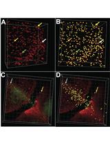
Looking through Brains with Fast Passive CLARITY: Zebrafish, Rodents, Non-human Primates and Humans

利用 Fast Passive CLARITY技术研究斑马鱼、啮齿动物、灵长类动物和人类的大脑

FM Farzad Mortazavi
AS Alexander J. Stankiewicz
IZ Irina V. Zhdanova
8498 Views
Aug 5, 2019
Recently developed CLARITY (Clear Lipid-exchanged Acrylamide-hybridized Rigid Imaging/Immunostaining/In situ-hybridization-compatible Tis-sue-hYdrogel) technique renders the tissue transparent by removing lipids in the tissue, while preserving and stabilizing the cellular and subcellular structures. This provides effective penetration of diverse labeling probes, from primary and secondary antibodies to complementary DNA and RNA strands. Followed by high-resolution 3D imaging of neuronal cells and their projections in thick sections, tissue blocks, whole brains, or whole animals, CLARITY allows for superior quantitative analysis of neuronal tissue. Here, we provide our detailed protocol for PACT (Passive Clarity Technique) in brain tissue of diverse species, including human, non-human primate, rodents, and zebrafish. We describe the six principal steps: (1) Tissue fixation and preparation, (2) Passive lipid removal, (3) Immuno-labeling, (4) Optical clearing, (5) Imaging, (6) 3D visualization and quantification.
"自由活动小鼠下丘脑区域间质液中前列腺素E2浓度的定量分析

Quantification of Prostaglandin E2 Concentration in Interstitial Fluid from the Hypothalamic Region of Free-moving Mice

"自由活动小鼠下丘脑区域间质液中前列腺素E2浓度的定量分析

YN Yoshinobu Nakamura
TS Takatoshi Sakaguchi
IT Ikumi Tamai
TN Takeo Nakanishi
4979 Views
Aug 5, 2019
Prostaglandin E2 (PGE2) is a well-established chemical mediator for the generation of the fever at the hypothalamus of the brain. PGE2 mediates fever generation via PGE receptor 3 (i.e., EP3) on neurons in the preoptic area. The role of PGE2 has been analyzed by measuring PGE2 concentration in cerebrospinal fluid (Ccsf); however, local PGE2 concentration at the hypothalamus may not necessarily be consistent with Ccsf. In this protocol, we introduce our method to measure directly the alteration in PGE2 concentration in interstitial fluid in the hypothalamus (Cisf) of awake (free-moving) mice using a microdialysis technique. Male mice (c57BL/6J) were anesthetized and fixed in the stereotaxic instrument, and a microdialysis probe was inserted into the hypothalamus through a guide cannula. On the fifth postoperative day, Cisf was monitored in free-moving mice that were intraperitoneally (i.p.) injected with lipopolysaccharide (LPS). PGE2 and other eicosanoids recovered in Krebs-Ringer phosphate buffer and defused through a microdialysis probe were extracted into ethyl acetate/formic acid and then quantified with LC-MS/MS. Our method is useful to understand the role of key regulators of prostaglandin concentration such as those of transporters, which have been unappreciated in inflammation-based brain diseases.

植物科学

拉伸强度测试用于检测拟南芥花序轴组织刚度

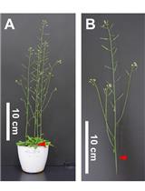
Tensile Testing Assay for the Measurement of Tissue Stiffness in Arabidopsis Inflorescence Stem

拉伸强度测试用于检测拟南芥花序轴组织刚度

KY Kouki Yoshida
SS Shingo Sakamoto
NM Nobutaka Mitsuda
9460 Views
Aug 5, 2019
Lignocellulosic biomass is a versatile renewable resource for fuels, buildings, crafts, and biomaterials. Strategies of molecularly designing lignocellulose for industrial application has been developed by the discoveries of novel genes after the screenings of various mutants and transformed lines of Arabidopsis whose cell walls could be modified in the inflorescence stem, a model woody tissue. The mechanical properties are used as a quantitative index for the chemorehological behavior of the genetically modified cell wall in the tissue. This parameter can be measured with tensile or bending tests of tissue explants, the vibration analysis of tissue behavior or using atomic force microscopy to probe the tissue surface. Here, we describe in detail the procedure to determine the stiffness of methanol-fixed, rehydrated and pronase-treated inflorescence explants with a tensile testing machine based on classical methods for the determination of cell wall extensibility.
拟南芥蛋白半衰期的检测

Measuring Protein Half-life in Arabidopsis thaliana

拟南芥蛋白半衰期的检测

Hongqing  Guo Hongqing Guo
Yanhai Yin Yanhai Yin
5282 Views
Aug 5, 2019
Post-translational modifications play important roles in controlling protein function and can lead to altered protein stability. Protein stability can be determined after treatment with the protein synthesis inhibitor Cycloheximide. Cycloheximide is a translational inhibitor that inhibits protein synthesis via cytoplasmic ribosomes. Here we describe how to measure the stability of MYC2 in the context of regulation by FERONIA receptor kinase. First, we describe how to measure MYC2 stability in wild-type and feronia mutant; then we describe similar assays in transgenic plants expressing MYC2-FLAG and MYC2A12-FLAG (12 FERONIA phosphorylation sites are mutated to Alanine and the mutant protein is stabilized). MYC2 can be induced by mechanical touch, which can be a confounding factor in protein level measurement. In this protocol, we take that into consideration and try to achieve more accurate measurement.

更正

更正:使用体外脉冲树突状细胞体外致敏T细胞: 腘窝淋巴结实验

Correction Notice: In vivo Priming of T Cells with in vitro Pulsed Dendritic Cells: Popliteal Lymph Node Assay

更正:使用体外脉冲树突状细胞体外致敏T细胞: 腘窝淋巴结实验

Songjie Cai Songjie Cai
MF Masayuki Fujino
LL Lina Lu
XL Xiao-Kang Li
1903 Views
Aug 5, 2019