往期刊物2019

卷册: 9, 期号: 5

left

Dec

20

Dec

5

Nov

20

Nov

5

Oct

20

Oct

5

Sep

20

Sep

5

Aug

20

Aug

5

Jul

20

Jul

5

Jun

20

Jun

5

May

20

May

5

Apr

20

Apr

5

Mar

20

Mar

5

Feb

20

Feb

5

Jan

20

Jan

5

right

生物化学

一种非放射性同位素寡糖基转移酶测定方法

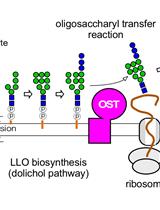
A Radioisotope-free Oligosaccharyltransferase Assay Method

一种非放射性同位素寡糖基转移酶测定方法

TY Takahiro Yamasaki
Daisuke Kohda Daisuke Kohda
5288 Views
Mar 5, 2019
Glycosylation of asparagine residues is widespread in Eukarya, and occurs in virtually all Archaea and some eubacterial species. A membrane-bound enzyme, oligosaccharyltransferase, catalyzes the transfer of an oligosaccharide chain from a sugar donor (lipid-linked oligosaccharide, LLO) to an asparagine residue in the consensus sequence, Asn-X-Ser/Thr (X ≠ Pro), in proteins. The in vitro oligosaccharyl transfer assay reaction mixture contains a detergent-solubilized oligosaccharyltransferase (OST), a sugar donor LLO, and a sugar acceptor peptide. Previous assay methods are problematic, in terms of the use of radioactive compounds and the cumbersome separation procedures using lectin binding or two-phase partitioning. Here, we describe a new oligosaccharyl transfer assay method, which is radioisotope-free and relies on a different separation mechanism. The glycopeptide products are separated from unreacted peptides by SDS-PAGE. A fluorescent dye is attached to the peptide substrate during custom peptide synthesis. The fluorescent imaging of the SDS-PAGE gels ensures high sensitivity and quantitative performance. The user-friendly PAGE format is particularly suitable for presentation in scientific papers. For illustrative applications, time-course and peptide library experiments are shown.

细胞生物学

人类精子的单细胞运动分析

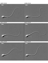
Single-cell Motility Analysis of Tethered Human Spermatozoa

人类精子的单细胞运动分析

WS William M. Skinner
NM Nadja Mannowetz
PL Polina V. Lishko
NR Nadia R. Roan
7033 Views
Mar 5, 2019
Vigorous sperm flagellar motility is essential for fertilization, and so the quantitative measurement of motility is a useful tool to assess the intrinsic fertility potential of sperm cells and explore how various factors can alter sperm’s ability to reach the egg and penetrate its protective layers. Human sperm beat their flagella many times each second, and so recording and accurately quantifying this movement requires a high-speed camera. The aim of this protocol is to provide a detailed description of the tools required for quantitative beat frequency measurement of tethered human sperm at the single-cell level and to describe methods for investigating the effects of intracellular or extracellular factors on flagellar motion. This assay complements bulk measurements of sperm parameters using commercially-available systems for computer-assisted sperm analysis (CASA).
一种直接离体测量人体表皮活检耗氧率的新方法

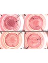
A New Efficient Method for Measuring Oxygen Consumption Rate Directly ex vivo in Human Epidermal Biopsies

一种直接离体测量人体表皮活检耗氧率的新方法

DS Daniel Schniertshauer
DG Daniel Gebhard
JB Jörg Bergemann
6315 Views
Mar 5, 2019
Skin cells are constantly exposed to environmental influences such as air pollution, chemicals, pathogens and UV radiation. UV radiation can damage different biological structures, but most importantly cellular DNA. Mitochondria contain their own genome and accumulate UV-induced DNA mutations to a large extent. This can result, e.g., in accelerated skin aging. Understanding the impact of harmful external influences on mitochondrial function is therefore essential for a better view on the development of age-related diseases. Previous studies have been carried out on cell cultures derived from primary cells, which does not fully represent the real situation in the skin, while the mitochondrial parameters were considered barely or not at all. Here we describe a method to measure mitochondrial respiratory parameters in epithelial tissue derived from human skin biopsies using an Agilent Seahorse XF24 Flux Analyzer. Before the assay, epidermis and dermis are separated enzymatically, we then used the XF24 Islet capture microplates to position the epidermis samples to measure oxygen consumption rates (OCR) and extracellular acidification rates (ECAR). In these plates, small nets can be fixed to the plate bottom. The epidermis was placed with the vital–basal–side on the net. Active ingredients in the three ports were injected consecutively to determine the effect of each compound. This allows determining the efficiency of the individual complexes within the respiratory chain. This protocol enables the testing of toxic substances and their influence on the mitochondrial respiration parameters in human epithelial tissue.

发育生物学

一种改进的可用于检测小鼠胚胎弱 LacZ 报告信号的染色法

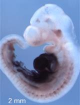
An Improved Staining Method for Low Signal LacZ Reporter Detection in Mouse Embryos

一种改进的可用于检测小鼠胚胎弱 LacZ 报告信号的染色法

XS Xiaopeng Shen
FX Feng Xu
SW Shen Wu
ML Meng Li
JZ Jingyi Zhang
RL Rui Liang
YL Yu Liu
6096 Views
Mar 5, 2019
In many fields of biology, especially in the field of developmental biology, LacZ reporter staining is an approach used to monitor gene expression patterns. In the LacZ reporter system, the LacZ gene is inserted in the endogenous location of the target gene via gene knock-in or by constructing a transgenic cassette in which LacZ is placed downstream of the promoter of the target gene being examined. Currently, the most common LacZ staining methods used are X-gal/FeCN staining and S-gal/TNBT staining. A serious limitation of both of these methods is that they are not effective when the LacZ gene is expressed at a low level. In an attempt to remedy this problem, we have established a new staining protocol which combines both methods. When compared to them, the method described here is better for visualizing lowly expressed genes and it has low background with high sensitivity.

免疫学

免疫荧光成像和流式细胞术鉴定小鼠成人睾丸巨噬细胞群体

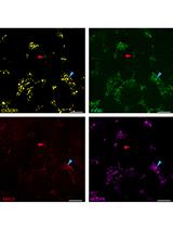
Characterization of Mouse Adult Testicular Macrophage Populations by Immunofluorescence Imaging and Flow Cytometry

免疫荧光成像和流式细胞术鉴定小鼠成人睾丸巨噬细胞群体

Noushin  Mossadegh-Keller Noushin Mossadegh-Keller
MS Michael H. Sieweke
6185 Views
Mar 5, 2019
Testicular macrophages (tMΦ) are the most abundant immune cells residing in the testis, an immune-privileged organ. TMΦ are known to exhibit different functions, such as protecting spermatozoa from auto-immune attack by producing immunosuppressive cytokines and trophic roles in supporting spermatogenesis and male sex hormone production. They also contribute to fetal testicular development. Recently, we characterized two distinct tMΦ populations based on their morphology, localization, cell surface markers, and gene expression profiling. Here, we focus and describe in detail the phenotypical distinction of these two tMΦ populations by fluorescence-activated cell sorting (FACS) using multicolor panel antibodies combining with high-resolution immunofluorescence (IF) imaging. These two techniques enable to classify two tMΦ populations: interstitial tMΦ and peritubular tMΦ.
BALB/c小鼠模型中早期卵白蛋白致敏和气溶胶攻击诱导的过敏性气道炎症

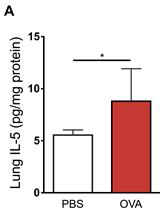
Early Life Ovalbumin Sensitization and Aerosol Challenge for the Induction of Allergic Airway Inflammation in a BALB/c Murine Model

BALB/c小鼠模型中早期卵白蛋白致敏和气溶胶攻击诱导的过敏性气道炎症

KM Kyle T Mincham
NS Naomi M Scott
JL Jean-Francois Lauzon-Joset
JL Jonatan Leffler
PS Philip A Stumbles
PH Patrick G Holt
DS Deborah H Strickland
5822 Views
Mar 5, 2019
The early life period represents a time of immunological plasticity whereby the functionally immature immune system is highly susceptible to environmental stimulation. Perennial aeroallergen and respiratory viral infection induced sporadic episodes of lung inflammation during this temporal window represent major risk factors for initiation of allergic asthmatic disease. Murine models are widely used as an investigative tool to examine the pathophysiology of allergic asthma; however, models in current usage typically do not encapsulate the early life period which represents the time of maximal risk for disease inception in humans. To address this issue, this protocol adapted an experimental animal model of disease for sensitization to ovalbumin during the immediate post-weaning period beginning at 21 days of age. By initially sensitizing mice during this early life post-weaning period, researchers can more closely align experimental allergic airway disease models with the human age group most at risk for asthma development.
通过体内被动皮肤过敏试验定量血清卵清蛋白特异性免疫球蛋白E滴度

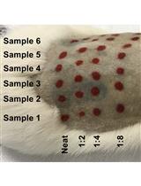
Quantification of Serum Ovalbumin-specific Immunoglobulin E Titre via in vivo Passive Cutaneous Anaphylaxis Assay

通过体内被动皮肤过敏试验定量血清卵清蛋白特异性免疫球蛋白E滴度

KM Kyle T Mincham
JL Jonatan Leffler
NS Naomi M Scott
JL Jean-Francois Lauzon-Joset
PS Philip A Stumbles
PH Patrick G Holt
DS Deborah Strickland
5436 Views
Mar 5, 2019
Murine models of allergic airway disease are frequently used as a tool to elucidate the cellular and molecular mechanisms of tissue-specific asthmatic disease pathogenesis. Paramount to the success of these models is the induction of experimental antigen sensitization, as indicated by the presence of antigen-specific serum immunoglobulin E. The quantification of antigen-specific serum IgE is routinely performed via enzyme-linked immunosorbent assay. However, the reproducibility of these in vitro assays can vary dramatically in our experience. Furthermore, quantifying IgE via in vitro methodologies does not enable the functional relevance of circulating IgE levels to be considered. As a biologically appropriate alternative method, we describe herein a highly reproducible in vivo passive cutaneous anaphylaxis assay using Sprague Dawley rats for the quantification of ovalbumin-specific IgE in serum samples from ovalbumin-sensitized murine models. Briefly, this in vivo assay involves subcutaneous injections of serum samples on the back of a Sprague Dawley rat, followed 24 h later by intravenous injection of ovalbumin and a blue detection dye. The subsequent result of antigen-IgE mediated inflammation and leakage of blue dye into the initial injection site indicates the presence of ovalbumin-specific IgE within the corresponding serum sample.

分子生物学

QsRNA-seq: 小RNA高通量测序文库构建方案

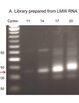
QsRNA-seq: A protocol for Generating Libraries for High-throughput Sequencing of Small RNAs

QsRNA-seq: 小RNA高通量测序文库构建方案

Alla Fishman Alla Fishman
Ayelet T. Lamm Ayelet T. Lamm
7512 Views
Mar 5, 2019
Small RNAs (sRNAs) are 20-30 nt long non-coding RNA molecules that regulate essentially all cellular processes. Besides being an intensively studied topic in academic research, sRNAs also hold a promise as clinical biomarkers. While the need for expressional profiling of sRNAs is growing, preparation of sRNA libraries for high-throughput sequencing (HTS) remains technically challenging, due to their small size. The common PAGE-based protocol is time-consuming and inefficient due to material loss, while gel-free protocols generate libraries of insufficient quality. To overcome these shortcomings, we modified the conditions of size-selection by Solid Phase Reversible Immobilization (SPRI) in a way that allows separation of nucleic acids shorter than 100 nt and differing in length by only 20 nt. Implementing the method for preparation of small RNA libraries for HTS resulted in QsRNA-seq, a gel-free, fast and easy-to-perform protocol, amenable to automation, generating very clean libraries that result in high-depth expression data. The protocol also utilizes Unique Molecular Identifiers (UMI) for reduction of library preparation biases and to quantify expression levels. QsRNA-seq provides an excellent solution to the growing needs for small RNA expression profiling for research clinical use.

神经科学

使用组织切碎机快速制备小脑切片

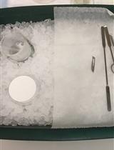
Acute Cerebellar Slice Preparation Using a Tissue Chopper

使用组织切碎机快速制备小脑切片

YW Yubin Wang
MB Michel Baudry
5276 Views
Mar 5, 2019
Acute cerebellar slices are widely used among neuroscientists to study the properties of excitatory and inhibitory synaptic transmission as well as intracellular signaling pathways involved in their regulation in cerebellum. The cerebellar cortex presents a well-organized circuitry, and several neuronal pathways can be stimulated and recorded reliably in acute cerebellar slices. A widely used acute cerebellar slice preparation technique was adapted from Edwards’ thin slice preparation method published in 1989 (Edwards et al., 1989). Most of the acute cerebellar slice preparation techniques use a vibrating microtome for slicing freshly dissected cerebellum from various animal species. Here we introduce a simpler method, which uses a tissue chopper to quickly prepare acute sagittal cerebellar slices from rodents. Cerebellum is dissected from the whole brain and sliced with a tissue chopper into 200-400 µm thick slices. Slices are allowed to recover in oxygenated aCSF at 37 °C for 1-2 h. Slices can then be used for electrophysiology or other types of experimentation. This method can be used to prepare cerebellar slices from mouse or rat aged from postnatal day 7 to 2 years. The preparation is faster and easier than other methods and provides a more versatile diversity of applications.

植物科学

单溴二甲双胍 (mBB) 检测拟南芥蛋白质提取物中的二硫化物

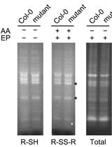
Detection of Disulfides in Protein Extracts of Arabidopsis thaliana Using Monobromobimane (mBB)

单溴二甲双胍 (mBB) 检测拟南芥蛋白质提取物中的二硫化物

SH Shin‐nosuke Hashida
MK Maki Kawai-Yamada
5709 Views
Mar 5, 2019
Thiol-disulfide exchange is a key posttranslational modification, determining the folding process of intra- and inter-protein structures. Thiols can be detected by colorimetric reagents, which are stoichiometrically reduced by free thiols, and by fluorescent adducts, showing fluorescence only after thioester formation. We adapted a simple three-step method for detection of disulfide bonds in proteins. After irreversible blocking of protein thiols, disulfide bonds are reduced, followed by the detection of thiols. The approach presented here provides an economical procedure that can be used to obtain a global overview of the thiol-disulfide status of proteins in plants. This method allows the detection of modifications in samples on a gel and can be used for semi-quantitative analysis.

干细胞

人诱导多能干细胞 (iPSCs) 分化为前脑神经祖细胞和成熟神经元的有效模型

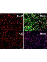
Differentiation of Human Induced Pluripotent Stem Cells (iPSCs) into an Effective Model of Forebrain Neural Progenitor Cells and Mature Neurons

人诱导多能干细胞 (iPSCs) 分化为前脑神经祖细胞和成熟神经元的有效模型

SB Scott Bell
NH Nuwan C. Hettige
HS Heika Silveira
HP Huashan Peng
HW Hanrong Wu
MJ Malvin Jefri
LA Lilit Antonyan
YZ Ying Zhang
XZ Xin Zhang
CE Carl Ernst
16349 Views
Mar 5, 2019
Induced Pluripotent Stem Cells (iPSCs) are pluripotent stem cells that can be generated from somatic cells, and provide a way to model the development of neural tissues in vitro. One particularly interesting application of iPSCs is the development of neurons analogous to those found in the human forebrain. Forebrain neurons play a central role in cognition and sensory processing, and deficits in forebrain neuronal activity contributes to a host of conditions, including epilepsy, Alzheimer’s disease, and schizophrenia. Here, we present our protocol for differentiating iPSCs into forebrain neural progenitor cells (NPCs) and neurons, whereby neural rosettes are generated from stem cells without dissociation and NPCs purified from rosettes based on their adhesion, resulting in a more rapid generation of pure NPC cultures. Neural progenitor cells can be maintained as long-term cultures, or differentiated into forebrain neurons. This protocol provides a simplified and fast methodology of generating forebrain NPCs and neurons, and enables researchers to generate effective in vitro models to study forebrain disease and neurodevelopment. This protocol can also be easily adapted to generate other neural lineages.