往期刊物2018

卷册: 8, 期号: 20

left

Dec

20

Dec

5

Nov

20

Nov

5

Oct

20

Oct

5

Sep

20

Sep

5

Aug

20

Aug

5

Jul

20

Jul

5

Jun

20

Jun

5

May

20

May

5

Apr

20

Apr

5

Mar

20

Mar

5

Feb

20

Feb

5

Jan

20

Jan

5

right

生物化学

秀丽隐杆线虫中细胞核/细胞质蛋白的分离

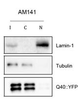
Nuclear/Cytoplasmic Fractionation of Proteins from Caenorhabditis elegans

秀丽隐杆线虫中细胞核/细胞质蛋白的分离

AM Alejandro Mata-Cabana
Olga Sin Olga Sin
RS Renée I. Seinstra
EN Ellen A. A. Nollen
10109 Views
Oct 20, 2018
C. elegans is widely used to investigate biological processes related to health and disease. To study protein localization, fluorescently-tagged proteins can be used in vivo or immunohistochemistry can be performed in whole worms. Here, we describe a technique to localize a protein of interest at a subcellular level in C. elegans lysates, which can give insight into the location, function and/or toxicity of proteins.

细胞生物学

小鼠药代动力学研究

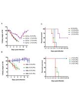
Murine Pharmacokinetic Studies

小鼠药代动力学研究

Alix F. Leblanc Alix F. Leblanc
Kevin M. Huang Kevin M. Huang
Muhammad Erfan Uddin Muhammad Erfan Uddin
JA Jason T. Anderson
MC Mingqing Chen
SH Shuiying Hu
14841 Views
Oct 20, 2018
Murine pharmacokinetics (PK) represents the absorption, distribution, metabolism, and elimination of drugs from the body, which helps to guide clinical studies, ultimately resulting in more effective drug treatment. The purpose of this protocol is to describe a serial bleeding protocol, obtaining blood samples at six time points from single mouse to yield a complete PK profile. This protocol has proved to be rapid, highly repeatable, and relatively easy to acquire. Comparing with the conventional PK studies, this method not only dramatically reduces animal usage, but also decreases sample variation obtained from different animals.
MDCK-II细胞间鞘糖脂运输的跨细胞转运检测

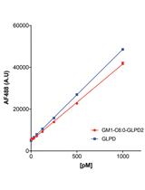
Transcytosis Assay for Transport of Glycosphingolipids across MDCK-II Cells

MDCK-II细胞间鞘糖脂运输的跨细胞转运检测

MG Maria Daniela Garcia-Castillo
Wayne I. Lencer Wayne I. Lencer
Daniel J.-F. Chinnapen Daniel J.-F. Chinnapen
7227 Views
Oct 20, 2018
Absorption and secretion of peptide and protein cargoes across single-cell thick mucosal and endothelial barriers occurs by active endocytic and vesicular trafficking that connects one side of the epithelial or endothelial cell (the lumen) with the other (the serosa or blood). Assays that assess this pathway must robustly control for non-specific and passive solute flux through weak or damaged intercellular junctions that seal the epithelial or endothelial cells together. Here we describe an in vitro cell culture Transwell assay for transcytosis of therapeutic peptides linked covalently to various species of the glycosphingolipid GM1. We recently used this assay to develop technology that harnesses endogenous mechanism of lipid sorting across epithelial cell barriers to enable oral delivery of peptide and protein therapeutics.
人冠状动脉和鼠头臂动脉的血管渗透性检测

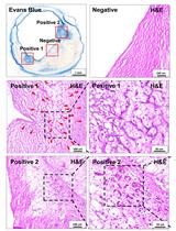
Vascular Permeability Assay in Human Coronary and Mouse Brachiocephalic Arteries

人冠状动脉和鼠头臂动脉的血管渗透性检测

LG Liang Guo
RF Raquel Fernandez
AS Atsushi Sakamoto
AC Anne Cornelissen
KP Ka Hyun Paek
PL Parker J. Lee
LW Leah M. Weinstein
CC Carlos J. Collado-Rivera
EH Emanuel Harari
RK Robert Kutys
TS Torie S. Samuda
NS Nicole A. Singer
MK Matthew D. Kutyna
FK Frank D. Kolodgie
RV Renu Virmani
AF Aloke V. Finn
6827 Views
Oct 20, 2018
Coronary artery disease remains an important cause of morbidity and mortality. Previous work, including ours, has focused on the role of intraplaque hemorrhage, particularly from immature microvessel angiogenesis, as an important contributor to plaque progression via increases in vascular permeability leading to further intraplaque hemorrhage, which increases red cell membrane-derived free cholesterol in plaque content and inflammatory cell recruitment. Evans Blue Dye (EBD) assay is widely used as a standard assay for vasculature permeability. However, the method has not been established in fresh human coronary artery autopsy samples to evaluate intraplaque microvessel permeability and angiogenesis. In this protocol, we describe a method to evaluate human coronary samples for microvascular permeability, including procedures to perfuse coronary arteries, collection of artery samples for histological analysis and immunostaining as well as the use of appropriate methodology to analyze the images. An optional procedure is also provided for the use of FITC-dextran in mouse model to evaluate vascular permeability. These Evans Blue Dye procedures may be useful in providing functional measure of the endothelium integrity and permeability in both human samples and animal models in various pathological conditions.

免疫学

一种体外检测cGAS活性的高效阴离子交换色谱法

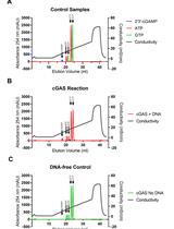
A Highly Sensitive Anion Exchange Chromatography Method for Measuring cGAS Activity in vitro

一种体外检测cGAS活性的高效阴离子交换色谱法

AH Andreas Holleufer
RH Rune Hartmann
5720 Views
Oct 20, 2018
Cyclic GMP-AMP synthase (cGAS) is a pattern recognition receptor (PRR) that senses double stranded DNA (dsDNA) in the cytosol and this leads to the activation of stimulator of interferon genes (STING) via the secondary messenger 2’3’-cyclic GMP-AMP (2’3’-cGAMP). STING then recruits TANK binding kinase 1 (TBK-1) and this complex can phosphorylate and activate interferon regulatory factor 3 (IRF3) leading to the induction of type I interferons and other antiviral genes. The cGAS:DNA complex catalyzes the synthesis of 2’3’-cGAMP and the purpose of the protocol presented here is to measure the in vitro activity of purified cGAS in the presence of dsDNA. The protocol was developed to elucidate the relationship between dsDNA length and the level of cGAS activity. The method involves an in vitro reaction with low concentrations of cGAS and dsDNA followed by quantification of the reaction product using anion exchange chromatography. The low concentrations of cGAS and dsDNA and the high sensitivity of this assay is a key advantage when comparing different DNA fragments’ ability to activate cGAS.

微生物学

利用荧光膜染料评估膜流动性以及观察细菌中的流体膜结构区域

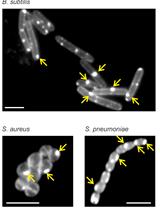
Assessing Membrane Fluidity and Visualizing Fluid Membrane Domains in Bacteria Using Fluorescent Membrane Dyes

利用荧光膜染料评估膜流动性以及观察细菌中的流体膜结构区域

Michaela Wenzel Michaela Wenzel
NV Norbert O. E. Vischer
HS Henrik Strahl
LH Leendert W. Hamoen
12536 Views
Oct 20, 2018
Membrane fluidity is a key parameter of bacterial membranes that undergoes quick adaptation in response to environmental challenges and has recently emerged as an important factor in the antibacterial mechanism of membrane-targeting antibiotics. The specific level of membrane fluidity is not uniform across the bacterial cell membrane. Rather, specialized microdomains associated with different cellular functions can exhibit fluidity values that significantly deviate from the average. Assessing changes in the overall membrane fluidity and formation of membrane microdomains is therefore pivotal to understand both the functional organization of the bacterial cell membrane as well as antibiotic mechanisms. Here we describe how two fluorescent membrane dyes, laurdan and DiIC12, can be employed to assess membrane fluidity in living bacteria. We focus on Bacillus subtilis, since this organism has been relatively well-studied with respect to membrane domains. However, we also describe how these assays can be adapted for other bacteria such as Staphylococcus aureus and Streptococcus pneumoniae.
HIVGKO:一种用于检测人原代CD4+T细胞中艾滋病潜伏期逆转剂的工具

HIVGKO: A Tool to Assess HIV-1 Latency Reversal Agents in Human Primary CD4+ T Cells

HIVGKO:一种用于检测人原代CD4+T细胞中艾滋病潜伏期逆转剂的工具

EB Emilie Battivelli
EV Eric Verdin
7527 Views
Oct 20, 2018
While able to suppress HIV replication in HIV infected individuals, combination antiretroviral therapy (ART) fails to eliminate viral latent reservoir, which consists in integrated transcriptional silenced HIV provirus. So far, identification of latently-infected cells has relied on activating cells to induce expression of HIV proteins which can then be detected. Unfortunately, this activation significantly changed the cell phenotype. We developed a novel HIV reporter, named HIVGKO, that allows the purification of latently-infected cells in absence of reactivation. Indeed, latent cells can be identified by expression of the EF1a-driven mKO2 and lack of expression of the LTR-driven csGFP. This protocol can be used to study the effectiveness of LRAs (Latency Reversal Agents) in reactivating latent HIV in primary cells.
H1N1病毒的制备和感染

H1N1 Virus Production and Infection

H1N1病毒的制备和感染

BZ Binbin Zhao
JS Jiaoyu Shan
RX Rui Xiong
KX Ke Xu
Bin Li Bin Li
6518 Views
Oct 20, 2018
Influenza A virus is a member of orthomyxoviridae family causing wide-spread infections in human respiratory tract. Mouse infection model is widely used in antiviral research and pathogenesis study against influenza A virus. Here, we report a protocol in infected mice with different virus doses and strains to explore how an inhibitor of lysine-specific demethylase (LSD1) impacts disease progression.
大豆疫霉菌分泌蛋白的制备和纯化

Preparation and Purification of Proteins Secreted from Phytophthora sojae

大豆疫霉菌分泌蛋白的制备和纯化

YX Yeqiang Xia
YW Yan Wang
Yuanchao   Wang Yuanchao Wang
6434 Views
Oct 20, 2018
Phytophthora sojae, the causal agent of soybean root and stem rot, is responsible for enormous economic losses in soybean production. P. sojae secrets various effectors to reprogram host immunity. The plant apoplastic space is a major battleground in plant-pathogen interactions. Here we describe a protocol for purification and isolation of secreted proteins from P. sojae, including precipitation of secreted proteins from P. sojae culture filtrate, chromatographic purification of the secreted proteins and analysis of the proteins by Mass spectrometry. With this protocol, it will be easier to identify potential apoplastic effectors in Phytophthora and will benefit our understanding of plant-microbe interactions.
分支DNA FISH技术联合共聚焦显微镜或流式细胞术检测和鉴别多样的病毒RNA

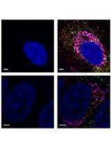
Detection and Differentiation of Multiple Viral RNAs Using Branched DNA FISH Coupled to Confocal Microscopy and Flow Cytometry

分支DNA FISH技术联合共聚焦显微镜或流式细胞术检测和鉴别多样的病毒RNA

Nv Nicholas van Buuren
KK Karla Kirkegaard
6001 Views
Oct 20, 2018
Due to the exceptionally high mutation rates of RNA-dependent RNA polymerases, infectious RNA viruses generate extensive sequence diversity, leading to some of the lowest barriers to the development of antiviral drug resistance in the microbial world. We have previously discovered that higher barriers to the development of drug resistance can be achieved through dominant suppression of drug-resistant viruses by their drug-susceptible parents. We have explored the existence of dominant drug targets in poliovirus, dengue virus and hepatitis C virus (HCV). The low replication capacity of HCV required the development of novel strategies for identifying cells co-infected with drug-susceptible and drug-resistant strains. To monitor co-infected cell populations, we generated codon-altered versions of the JFH1 strain of HCV. Then, we could differentiate the codon-altered and wild-type strains using a novel type of RNA fluorescent in situ hybridization (FISH) coupled with flow cytometry or confocal microscopy. Both of these techniques can be used in conjunction with standard antibody-protein detection methods. Here, we describe a detailed protocol for both RNA FISH flow cytometry and confocal microscopy.
稻株中水稻齿叶矮缩病毒的传播和感染

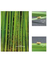
Rice Ragged Stunt Virus Propagation and Infection on Rice Plants

稻株中水稻齿叶矮缩病毒的传播和感染

CZ Chao Zhang
CS Chaonan Shi
DC Dong Chen
Jianguo   Wu Jianguo Wu
5699 Views
Oct 20, 2018
Virus inoculation is a basic experimental procedure to evaluate the resistance of a rice variety or a transgenic material upon virus infection. We recently demonstrated that Rice Ragged Stunt Virus (RRSV), an oryzavirus that is transmitted by brown planthopper (BPH), can suppress jasmonic acid-mediated antiviral defense through the induction of microRNA319 and facilitate virus infection in rice. To verify this, we performed virus inoculation experiments on wild-type rice plants and miR319-TCP21-associated transgenic rice plants through a modified group inoculation method. Here, we presented the detailed procedure of RRSV propagation and infection process on rice plants.

分子生物学

通过CRISPR-Trap完全移除内源全长转录本进行基因敲除和基因置换

Generation of Gene Knockout and Gene Replacement with Complete Removal of Full-length Endogenous Transcript Using CRISPR-Trap

通过CRISPR-Trap完全移除内源全长转录本进行基因敲除和基因置换

JM Jonas Mechtersheimer
Stefan Reber Stefan Reber
Marc-David Ruepp Marc-David Ruepp
8480 Views
Oct 20, 2018
This protocol describes the application of the CRISPR-Trap from designing of the gene targeting strategy to validation of successfully edited clones that was validated on various human cell lines, among them human induced pluripotent stem cells (hiPSCs). The advantage of CRISPR-Trap over conventional approaches is the complete removal of any endogenous full-length transcript from the target gene. CRISPR-Trap is applicable for any target gene with no or little coding sequence in its first exon. Several human cell lines and different genes have so far been edited successfully with CRISPR-Trap.
基于微量滴定板的Cell-SELEX技术

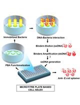
Microtitre Plate Based Cell-SELEX Method

基于微量滴定板的Cell-SELEX技术

MS Munish Shorie
HK Harmanjit Kaur
7457 Views
Oct 20, 2018
Aptamers have emerged as a novel category in the field of bioreceptors due to their wide applications ranging from biosensing to therapeutics. Several variations of their screening process, called SELEX have been reported which can yield sequences with desired properties needed for their final use. We report a facile microtiter plate-based Cell-SELEX method for a gram-negative bacteria E. coli. The optimized protocol allows the reduction of number of rounds for SELEX by offering higher surface area and longer retention times. In addition, this protocol can be modified for other prokaryotic and eukaryotic cells, and glycan moieties as target for generation of high affinity bio-receptors in a short course of time in-vitro.

神经科学

利用SeeD2培养基对组织样品光透明及折射率匹配以实现高分辨率成像

Optical Clearing and Index Matching of Tissue Samples for High-resolution Fluorescence Imaging Using SeeDB2

利用SeeD2培养基对组织样品光透明及折射率匹配以实现高分辨率成像

MK Meng-Tsen Ke
TI Takeshi Imai
12141 Views
Oct 20, 2018
Tissue clearing techniques are useful for large-scale three-dimensional fluorescence imaging of thick tissues. However, high-resolution imaging deep inside tissues has been challenging, as it is extremely sensitive to light scattering and spherical aberrations. Here, we present a water-based optical clearing and mounting media, SeeDB2, which is designed for high numerical aperture (NA) objective lenses with oil or glycerol immersion. Using quick and simple soaking procedures, the refractive indices of samples can be matched either to that of immersion oil (1.52) or glycerol (1.46), thus minimizing light scattering and spherical aberrations. Fine morphology and various fluorescent proteins are highly preserved during the clearing and imaging process. Our method is useful for the three-dimensional fluorescence imaging of neuronal circuitry at synaptic resolution using confocal and super-resolution microscopy. SeeDB2 is also useful as a mounting media for the super-resolution imaging of fluorescent proteins.
承受应激小鼠的觅食和酒精偏好的行为学评估

Behavioral Evaluation of Seeking and Preference of Alcohol in Mice Subjected to Stress

承受应激小鼠的觅食和酒精偏好的行为学评估

AC Ana Canseco-Alba
NS Norman Schanz
HI Hiroki Ishiguro
QL Qing-Rong Liu
Emmanuel S. Onaivi Emmanuel S. Onaivi
6058 Views
Oct 20, 2018
The alcohol preference model is one of the most widely used animal models relevant to alcoholism. Stressors increase alcohol consumption. Here we present a protocol for a rapid and useful tool to test alcohol preference and stress-induced alcohol consumption in mice. In this model, animals are given two bottles, one with a diluted solution of ethanol in water, and the other with tap water. Consumption from each bottle is monitored over a 24-h period over several days to assess the animal’s relative preference for the ethanol solution over water. In the second phase, animals are stressed by restraining them for an hour daily and their subsequent preference of tap water or the ethanol solution is evaluated. Preference is measured by the volume and/or weight or liquid consumed daily, which is then converted to a preference ratio. The alcohol preference model was combined with the conditioned place preference paradigm to determine alcohol conditioning and preference following the deletion of CB2 cannabinoid receptors in dopaminergic neurons in the DAT-Cnr2 Cre-recombinant conditional knockout (cKO) mice in comparison with the wild-type control mice.
通过饮食控制测定黑腹果蝇的选型交配

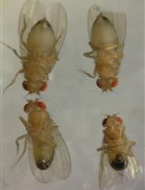
Testing for Assortative Mating by Diet in Drosophila melanogaster

通过饮食控制测定黑腹果蝇的选型交配

PL Philip T Leftwich
TC Tracey Chapman
5155 Views
Oct 20, 2018
Experimental studies of the evolution of reproductive isolation in real time are a powerful way to reveal the way that fundamental processes, such as mate choice, initiate divergence. Mate choice, while frequently described in females, can occur in either sex, and can be affected by the genetics or environment of an individual. Here we describe simple protocols for assessing mating outcomes in fruit flies, which in this context can be used to assess reproductive isolation derived from rearing on different diets over multiple generations.

植物科学

利用透射电镜检测植物细胞自噬

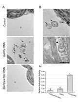
Examining Autophagy in Plant by Transmission Electron Microscopy (TEM)

利用透射电镜检测植物细胞自噬

XZ Xiyin Zheng
CZ Chenguang Zhao
Yule  Liu Yule Liu
7247 Views
Oct 20, 2018
In plants, macroautophagy, here referred as autophagy, is a degradation pathway during which the double-membrane structure named autophagosome engulfs the cargo and then fuses with vacuole for material recycling. To investigate the process of autophagy, transmission electron microscopy (TEM) was used to monitor the ultrastructure of autophagic structures and identify the cargo during this process due to its high resolution. Compared to other autophagy examination methods including biochemical assays and confocal microscopy, TEM is the only method that indicates the morphology of autophagic structures in nanoscale, which is considered to be one of the best ways to illustrate the morphology of autophagic intermediates and the substrate of autophagy. Here, we describe the autophagy examination assay using TEM in Nicotiana benthamiana leaf cells.
气动法检测植物木质部栓塞

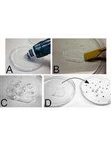
Pneumatic Method to Measure Plant Xylem Embolism

气动法检测植物木质部栓塞

PB Paulo R. L. Bittencourt
LP Luciano Pereira
RO Rafael S. Oliveira
6674 Views
Oct 20, 2018
Embolism, the formation of air bubbles in the plant water transport system, has a major impact on plant water relations. Embolism formation in the water transport system of plants disrupts plant water transport capacity, impairing plant functioning and triggering plant mortality. Measuring embolism with traditional hydraulic methods is both time-consuming and requires large amounts of plant material. While the stem hydraulic methods measure loss of xylem hydraulic conductance due to embolism formation, the pneumatic method directly quantifies the amount of emboli inside the xylem as changes in xylem air content. The pneumatic method is an easy and fast (8+ embolism curves per day) method to measure plant embolism requiring minimal plant material. Here, we provide detailed descriptions and recent technical improvements on the pneumatic method.