往期刊物2018

卷册: 8, 期号: 9

left

Dec

20

Dec

5

Nov

20

Nov

5

Oct

20

Oct

5

Sep

20

Sep

5

Aug

20

Aug

5

Jul

20

Jul

5

Jun

20

Jun

5

May

20

May

5

Apr

20

Apr

5

Mar

20

Mar

5

Feb

20

Feb

5

Jan

20

Jan

5

right

生物化学

长寿蛋白降解测定法:定量测定粘附细胞系中内源性蛋白自噬通量的有效方法

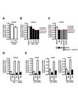
The Long-lived Protein Degradation Assay: an Efficient Method for Quantitative Determination of the Autophagic Flux of Endogenous Proteins in Adherent Cell Lines

长寿蛋白降解测定法:定量测定粘附细胞系中内源性蛋白自噬通量的有效方法

Morten  Luhr Morten Luhr
FS Frank Sætre
Nikolai   Engedal Nikolai Engedal
10266 Views
May 5, 2018
Autophagy is a key player in the maintenance of cellular homeostasis in eukaryotes, and numerous diseases, including cancer and neurodegenerative disorders, are associated with alterations in autophagy. The interest for studying autophagy has grown intensely in the last two decades, and so has the arsenal of methods utilised to study this highly dynamic and complex process. Changes in the expression and/or localisation of autophagy-related proteins are frequently assessed by Western blot and various microscopy techniques. Such analyses may be indicative of alterations in autophagy-related processes and informative about the specific marker being investigated. However, since these proteins are part of the autophagic machinery, and not autophagic cargo, they cannot be used to draw conclusions regarding autophagic cargo flux. Here, we provide a protocol to quantitatively assess bulk autophagic flux by employing the long-lived protein degradation assay. Our procedure, which traces the degradation of 14C valine-labelled proteins, is simple and quick, allows for processing of a relatively large number of samples in parallel, and can in principle be used with any adherent cell line. Most importantly, it enables quantitative measurements of endogenous cargo flux through the autophagic pathway. As such, it is one of the gold standards for studying autophagic activity.

癌症生物学

采用基于成像的分析法进行粘附细胞的细胞增殖和细胞死亡高通量分析

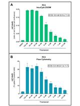
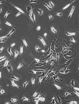
An Image-based Assay for High-throughput Analysis of Cell Proliferation and Cell Death of Adherent Cells

采用基于成像的分析法进行粘附细胞的细胞增殖和细胞死亡高通量分析

Paula  Szalai Paula Szalai
Nikolai  Engedal Nikolai Engedal
10168 Views
May 5, 2018
In this protocol, we describe a method to monitor cell proliferation and death by live-cell imaging of propidium iodide (PI)-stained adherent mammalian cells. PI is widely used to assess cell death. However, it is usually used in end-point assays. Recently, we implemented the use of PI for real-time cell death assessment by automated imaging. Cells are seeded in a 96-well format, and after attachment, the treatments are added directly to the wells together with PI. Thereafter, cells are subjected to automated time-lapse imaging and quantification by computer software. Combined analyses of phase-contrast and fluorescence images allow assessment of treatment effects on cell proliferation as well as the extent and kinetics of cell death.

细胞生物学

使用基于液体的细胞学检查和WT1免疫酶染色定量人尿中的足细胞和壁层上皮细胞

Quantifying Podocytes and Parietal Epithelial Cells in Human Urine Using Liquid-based Cytology and WT1 Immunoenzyme Staining

使用基于液体的细胞学检查和WT1免疫酶染色定量人尿中的足细胞和壁层上皮细胞

HO Hiroyuki Ohsaki
TM Toru Matsunaga
TF Taishi Fujita
YT Yasunori Tokuhara
SK Shingo Kamoshida
TS Tadashi Sofue
6957 Views
May 5, 2018
In glomerular disease, podocytes and parietal epithelial cells (PECs) are shed in the urine. Therefore, urinary podocytes and PECs are noninvasive biomarkers of glomerular disease. The purpose of this protocol is to employ immunocytochemistry to detect podocytes and PECs, using the WT1 antibody on liquid-based cytology slides.

免疫学

通过氯化锂沉淀法纯化DSS处理的鼠组织的总RNA

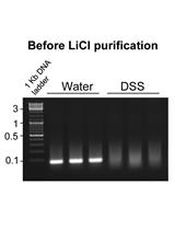
Purification of Total RNA from DSS-treated Murine Tissue via Lithium Chloride Precipitation

通过氯化锂沉淀法纯化DSS处理的鼠组织的总RNA

Emilie Viennois Emilie Viennois
AT Anika Tahsin
DM Didier Merlin
8749 Views
May 5, 2018
We have developed a protocol to purify RNA from DSS (Dextran Sulfate Sodium)-treated mouse tissues. This method, which prevents downstream inhibition of q-RT-PCR observed in DSS-treated tissues, relies on successive precipitations with lithium chloride.
从小鼠肌肉中分离Nab逃脱AAV突变体

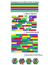
Nab Escaping AAV Mutants Isolated from Mouse Muscles

从小鼠肌肉中分离Nab逃脱AAV突变体

ZC Zheng Chai
RS R. Jude Samulski
CL Chengwen Li
6828 Views
May 5, 2018
Neutralizing antibodies (Nabs) are a major challenge in clinical trials of adeno-associated virus (AAV) vector gene therapy, because Nabs are able to inhibit AAV transduction in patients. We have successfully isolated several novel Nab-escaped AAV chimeric capsids in mice by administrating a mixture of AAV shuffled library and patient serum. These AAV chimeric capsid mutants enhanced Nab evasion from patient serum with a high muscle transduction efficacy. In this protocol, we describe the procedures for selection of the Nab-escaped AAV chimeric capsid, including isolation and characterization of Nab-escaping AAV mutants in mice muscle.

微生物学

CRISPR-Cas12a/Cpf1的异源表达和纯化

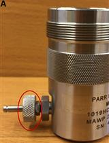
Heterologous Expression and Purification of the CRISPR-Cas12a/Cpf1 Protein

CRISPR-Cas12a/Cpf1的异源表达和纯化

PM Prarthana Mohanraju
John van der Oost John van der Oost
MJ Martin Jinek
Daan C. Swarts Daan C. Swarts
20558 Views
May 5, 2018
This protocol provides step by step instructions (Figure 1) for heterologous expression of Francisella novicida Cas12a (previously known as Cpf1) in Escherichia coli. It additionally includes a protocol for high-purity purification and briefly describes how activity assays can be performed. These protocols can also be used for purification of other Cas12a homologs and the purified proteins can be used for subsequent genome editing experiments. Figure 1. Timeline of activities for the heterologous expression and purification of Francisella novicida Cas12a (FnCas12a) from Escherichia coli
改变药物添加时间及变温分析实验确定抗病毒物质所作用的病毒生命周期中的步骤

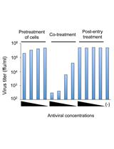
Time-of-addition and Temperature-shift Assays to Determine Particular Step(s) in the Viral Life Cycle that is Blocked by Antiviral Substance(s)

改变药物添加时间及变温分析实验确定抗病毒物质所作用的病毒生命周期中的步骤

CA Chie Aoki-Utsubo
MC Ming Chen
Hak Hotta Hak Hotta
9286 Views
May 5, 2018
Viruses infect their host cells to produce progeny virus particles through the sequential steps of the viral life cycle, such as viral attachment, entry, penetration and post-entry events. This protocol describes time-of-addition and temperature-shift assays that are employed to explore which step(s) in the viral life cycle is blocked by an antiviral substance(s).
大麦叶片上稻瘟病菌感染过程观察

Infection Process Observation of Magnaporthe oryzae on Barley Leaves

大麦叶片上稻瘟病菌感染过程观察

Xiao-Lin  Chen Xiao-Lin Chen
8993 Views
May 5, 2018
Rice blast and wheat blast caused by Magnaporthe oryzae is a serious threat to rice and wheat production. Appropriate methods for observing M. oryzae infection process are important for study of the fungal infection mechanisms, plant resistance reactions, and host-M. oryzae interactions. The rice leaf sheath is commonly used to inoculate M. oryzae for observing the infection process, however, this method is a time-consuming and high technical work. Here, we describe an easier solution to observe M. oryzae infection process on barley leaves.
蓝藻集胞藻PCC 6803中糖原和胞外葡萄糖估测

Glycogen and Extracellular Glucose Estimation from Cyanobacteria Synechocystis sp. PCC 6803

蓝藻集胞藻PCC 6803中糖原和胞外葡萄糖估测

MK Md. Rezaul Islam Khan
YW Yushu Wang
SA Shajia Afrin
LH Lin He
GM Gang Ma
6465 Views
May 5, 2018
Cyanobacteria, which have the extraordinary ability to grow using sunlight and carbon dioxide, are emerging as a green host to produce value-added products. Exploitation of this highly promising host to make products may depend on the ability to modulate the glucose metabolic pathway; it is the key metabolic pathway that generates intermediates that feed many industrially important pathways. Thus, before cyanobacteria can be considered as a leading source to produce value-added products, we must understand the interaction between glucose metabolism and other important cellular activities such as photosynthesis and chlorophyll metabolism. Here we describe reproducible and reliable methods for measuring extracellular glucose and glycogen levels from cyanobacteria.
通过免疫共沉淀法检测植物中双链RNA结合蛋白、病毒蛋白和初级miRNA转录产物的交互作用

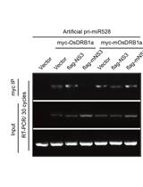
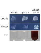
Detecting the Interaction of Double-stranded RNA Binding Protein, Viral Protein and Primary miRNA Transcript by Co-immunoprecipitation in planta

通过免疫共沉淀法检测植物中双链RNA结合蛋白、病毒蛋白和初级miRNA转录产物的交互作用

LZ Lijia Zheng
CZ Chao Zhang
Jianguo  Wu Jianguo Wu
YL Yi Li
6094 Views
May 5, 2018
MicroRNAs (miRNAs) play important roles in plant growth, development, and response to infection by microbes. Double-stranded RNA binding protein 1 (DRB1) facilitates the processing of primary miRNA transcripts into mature miRNAs. Recently, we found that NS3 protein encoded by rice stripe virus (RSV) associates with DRB1 and promotes miRNA biogenesis during RSV infection (Zheng et al., 2017). RNA co-immunoprecipitation (RIP) method was applied to identity association patterns among DRB1, NS3, and miRNA transcript.
小分子抑制剂在持续性诺如病毒感染小鼠模型中的疗效评估

Assessing the Efficacy of Small Molecule Inhibitors in a Mouse Model of Persistent Norovirus Infection

小分子抑制剂在持续性诺如病毒感染小鼠模型中的疗效评估

Jana Van Dycke Jana Van Dycke
Johan Neyts Johan Neyts
JR Joana Rocha-Pereira
5988 Views
May 5, 2018
Human norovirus is the most common cause of acute gastroenteritis worldwide, resulting in estimated mortality of ~210,000 each year, of whom most are children under the age of five. However, norovirus can infect people of all age groups. There is a risk of prolonged infection in children, the elderly and patients who are immunocompromised. To study the inhibition of persistent norovirus replication by small molecule antivirals in vivo, we used a murine norovirus CR6 strain (MNV.CR6). We demonstrated earlier that efficient small molecules can reduce viral shedding in the stool of infected mice. Here we present how to generate the MNV.CR6 virus stock, infect type I and II interferon receptor knockout AG129 mice via oral gavage, administer antivirals to mice, and quantify viral genome copies in the stool of these mice.
改良的低量 RNA-Seq法通过检测小亚基核糖体RNA以分析微生物群落和多样性

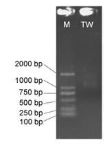
A Modified Low-quantity RNA-Seq Method for Microbial Community and Diversity Analysis Using Small Subunit Ribosomal RNA

改良的低量 RNA-Seq法通过检测小亚基核糖体RNA以分析微生物群落和多样性

YY Yong-Wei Yan
TZ Ting Zhu
BZ Bin Zou
Zhe-Xue Quan Zhe-Xue Quan
5571 Views
May 5, 2018
We propose a modified RNA-Seq method for small subunit ribosomal RNA (SSU rRNA)-based microbial community analysis that depends on the direct ligation of a 5’ adaptor to RNA before reverse-transcription. The method requires only a low-input quantity of RNA (10-100 ng) and does not require a DNA removal step. Using this method, we could obtain more 16S rRNA sequences of the same regions (variable regions V1-V2) without the interference of DNA in order to analyze OTU (operational taxonomic unit)-based microbial communities and diversity. The generated SSU rRNA sequences are also suitable for the coverage evaluation for bacterial universal primer 8F (Escherichia coli position 8 to 27), which is commonly used for bacterial 16S rRNA gene amplification. The modified RNA-Seq method will be useful to determine potentially active microbial community structures and diversity for various environmental samples, and will also be useful for identifying novel microbial taxa.

分子生物学

哺乳动物细胞源性囊泡用于分离细胞器特异性跨膜蛋白以进行单分子研究

Mammalian Cell-derived Vesicles for the Isolation of Organelle Specific Transmembrane Proteins to Conduct Single Molecule Studies

哺乳动物细胞源性囊泡用于分离细胞器特异性跨膜蛋白以进行单分子研究

FM Faruk H. Moonschi
AF Ashley M. Fox-Loe
XF Xu Fu
CR Chris I. Richards
6821 Views
May 5, 2018
Cell-derived vesicles facilitate the isolation of transmembrane proteins in their physiological membrane maintaining their structural and functional integrity. These vesicles can be generated from different cellular organelles producing, housing, or transporting the proteins. Combined with single-molecule imaging, isolated organelle specific vesicles can be employed to study the trafficking and assembly of the embedded proteins. Here we present a method for organelle specific single molecule imaging via isolation of ER and plasma membrane vesicles from HEK293T cells by employing OptiPrep gradients and nitrogen cavitation. The isolation was validated through Western blotting, and the isolated vesicles were used to perform single molecule studies of oligomeric receptor assembly.

植物科学

分泌蛋白信号肽功能评估

Functional Evaluation of the Signal Peptides of Secreted Proteins

分泌蛋白信号肽功能评估

Weixiao  Yin Weixiao Yin
YW Yufu Wang
TC Tao Chen
YL Yang Lin
CL Chaoxi Luo
11780 Views
May 5, 2018
Here, we describe a method that can be used to evaluate the function of predicted signal peptides. This method utilizes the yeast Saccharomyces cerevisiae YTK12 strain and pSUC2 vector in which the pSUC2 vector with fused predicted signal peptide is transformed into yeast. The function of the signal peptides can be evaluated by using different selective media and color reaction. In this protocol, we provide the detailed description of manipulation in order to implement easily.
通过测定蓝藻叶绿素荧光评估呼吸和光合作用的条件

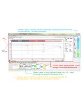
Evaluation of the Condition of Respiration and Photosynthesis by Measuring Chlorophyll Fluorescence in Cyanobacteria

通过测定蓝藻叶绿素荧光评估呼吸和光合作用的条件

TO Takako Ogawa
KS Kintake Sonoike
7598 Views
May 5, 2018
Chlorophyll fluorescence measurements have been widely used to monitor the condition of photosynthesis. Furthermore, chlorophyll fluorescence from cyanobacteria reflects the condition of respiration, since cyanobacterial photosynthesis shares several components of electron transport chain with respiration. This protocol presents the method to monitor the condition of both photosynthesis and respiration in cyanobacteria simply by measuring chlorophyll fluorescence in the dark and in the light with pulse amplitude modulation (PAM) chlorophyll fluorometer.

干细胞

小鼠骨髓造血干细胞的鹅卵石样区域形成细胞测定

Cobblestone Area-forming Cell Assay of Mouse Bone Marrow Hematopoietic Stem Cells

小鼠骨髓造血干细胞的鹅卵石样区域形成细胞测定

SA Surya Amarachintha
QP Qishen Pang
10067 Views
May 5, 2018
Bone Marrow Hematopoietic Stem Cells (HSCs) require bone marrow microenvironment for their maintenance and proliferation. Culture of Bone Marrow Mesenchymal Stromal Cells (MSCs) provides appropriate environmental signals for HSCs survival in vitro. Here, we provide a detailed protocol that describes culture conditions for MSCs, flow cytometric isolation of HSCs from mouse bone marrow, and perform co-culture of MSCs and HSCs known as Cobblestone area-forming cell (CAFC) assay. Altogether, CAFC assays can be used as a high-throughput in vitro screening model where efforts are made to understand and develop therapies for complex bone marrow diseases. This protocol needs 3 to 4 weeks starting from culturing MSCs, isolating LSK cells (HSCs), and to performing limited dilution CAFC assay.